
The Excretory System | ICSE Class 10 Biology Notes
On this page
Watch & explore
Start with a few high-quality watches, then dive into the notes below.
Try an idea before you read. Design a 2-step scenario interactive for a study note. Explore →

This note covers excretion, human excretory organs, the urinary system, external and internal kidney structure, renal blood vessels, the nephron, ultrafiltration, selective reabsorption, tubular secretion, urine composition and the passage of urine out of the body.
What is excretion, and which organs help remove wastes?
Excretion is the biological removal of harmful metabolic wastes from the body. Metabolism means the chemical activities occurring in living cells. These activities produce substances that must be removed rather than allowed to accumulate in body fluids.
Nitrogenous wastes are waste substances containing nitrogen. Ammonia, urea and uric acid are major examples. In humans, the kidneys remove nitrogenous wastes from the blood. Carbon dioxide, water and excess ions also need to be eliminated totally or partially.
An ion is an electrically charged particle. Examples include sodium, potassium and chloride ions. Removal of these substances is linked with maintaining an appropriate composition of body fluids, rather than simply removing every substance that enters the kidneys.
How do the liver, lungs and skin contribute?
The liver converts ammonia produced by metabolism into urea, a less toxic nitrogenous waste. Urea enters the blood and reaches the kidneys for removal. Thus, the site where urea is formed differs from the organs that chiefly eliminate it.
The lungs remove carbon dioxide and significant quantities of water. The liver also eliminates substances through bile, its digestive secretion. These include bilirubin and biliverdin, which are bile pigments, the colouring substances in bile, as well as cholesterol, a fatty substance, and some drugs. Most of these substances ultimately leave with digestive wastes.
Sweat glands in the skin produce a watery secretion containing sodium chloride, small amounts of urea and lactic acid, an organic acid. Sweat primarily helps cool the body surface, but it also removes some wastes. Sweating is therefore an additional route of excretion.
Sebaceous glands, the oil-producing glands of the skin, eliminate certain substances through sebum, their oily secretion. Sebum includes substances such as waxes and provides a protective oily covering. Its function differs from the cooling effect of sweat.
Definition: Excretion removes metabolic wastes. Egestion is the removal of undigested food from the digestive tract. The two terms describe different materials, even though some excretory substances leave the body along with digestive wastes.
The kidneys, lungs, liver and skin consequently contribute in different ways. Naming an organ should be accompanied by identifying the material it removes. This prevents the mistaken idea that every excretory organ produces urine or that all waste removal occurs through the urinary system.
What are the parts of the human urinary system?
The urinary system consists of a pair of kidneys, a pair of ureters, a urinary bladder and a urethra. Each part has a distinct role in forming urine, transporting it, storing it or releasing it from the body.
The kidneys are situated in the abdomen, one on either side of the backbone. They remove nitrogenous wastes from blood and form urine. The ureters are the tubes that carry urine from the kidneys to the urinary bladder.
The urinary bladder is a muscular organ that stores urine. The urethra is the passage through which urine leaves the bladder and the body. A ureter and the urethra therefore connect different places and should not be given interchangeable labels.
How do blood vessels and urinary passages differ?
The word renal means relating to the kidney. The renal artery carries blood towards the kidney, while the renal vein carries blood away from it. These vessels carry blood, whereas the ureter carries urine formed within the kidney.
| Structure | Material carried or stored | Main role |
|---|---|---|
| Kidney | Blood is processed and urine is formed | Removes wastes and adjusts water and dissolved substances |
| Renal artery | Blood travelling towards the kidney | Brings blood into the kidney |
| Renal vein | Blood travelling away from the kidney | Carries blood away after passage through the kidney |
| Ureter | Urine | Connects a kidney with the urinary bladder |
| Urinary bladder | Urine | Stores urine before its release |
| Urethra | Urine | Conducts urine out of the bladder and body |
What the figure shows
Human urinary system
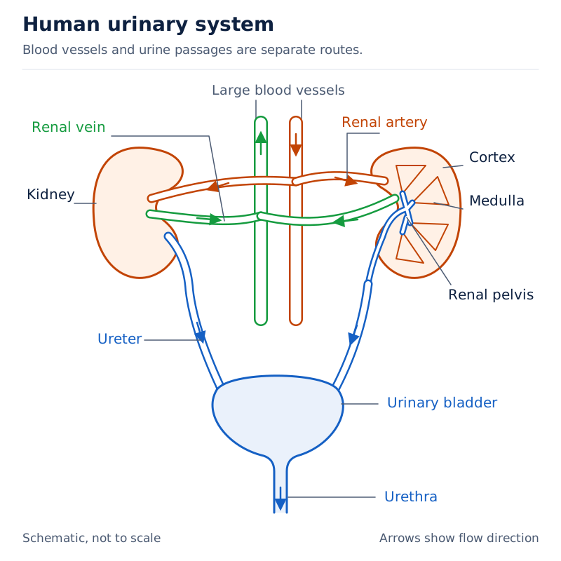
Two kidneys lie beside the central large blood vessels. Renal arteries and veins connect them with these vessels. Two ureters descend to the urinary bladder, and the urethra continues below it. One kidney is shown in section with cortex, medulla and pelvis labelled.
Drawn by One Young India.
The cortex is the kidney's outer internal zone, the medulla its inner zone, and the renal pelvis the funnel-shaped space that receives urine within the kidney. These labels belong to kidney structure, rather than to separate organs outside it.
Follow two routes when reading this diagram: blood enters and leaves through blood vessels; urine passes from the kidneys towards the bladder. Keeping these routes separate explains why the renal vein cannot replace the ureter as the path by which urine leaves a kidney.
How is a kidney organised externally and internally?
A kidney is a reddish-brown, bean-shaped organ close to the inner back wall of the abdominal cavity. Its inner surface is concave, meaning curved inwards. Towards the centre of this surface is a notch called the hilum, associated with the ureter, blood vessels and nerves.
A tough outer renal capsule covers the kidney. Beneath this covering are the cortex and medulla. The renal capsule surrounding the whole kidney must be distinguished from Bowman's capsule, the cup-like beginning of an individual kidney tubule.
What does a section through the kidney show?
The medulla is divided into conical masses called medullary pyramids. They project into cup-like extensions of the renal pelvis called calyces; one such extension is a calyx. The cortex extends between adjacent pyramids as renal columns, also called columns of Bertini.
The renal pelvis lies inside the hilum. It is continuous with the route leading into the ureter. This arrangement connects the tiny urine-forming structures inside the kidney with the tube that transports urine to the bladder.
| Part | Position or appearance |
|---|---|
| Renal capsule | Tough outer covering of the kidney |
| Cortex | Outer internal zone beneath the capsule |
| Medulla | Inner zone arranged into medullary pyramids |
| Renal columns | Extensions of cortex between the pyramids |
| Calyces | Extensions of the renal pelvis receiving the projecting pyramids |
| Renal pelvis | Broad funnel-shaped space internal to the hilum |
What the figure shows
Longitudinal section of a kidney
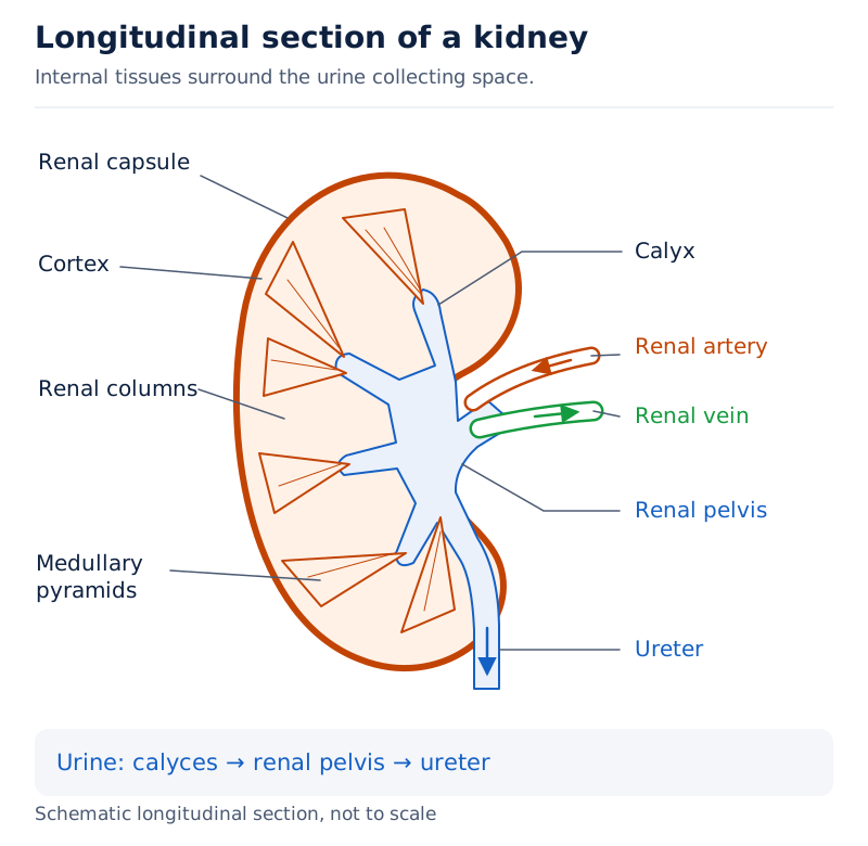
The cut surface shows the renal capsule and cortex around the outside, with medullary pyramids further in. Renal columns lie between pyramids. A calyx, the renal pelvis and the ureter are labelled, together with the renal artery and renal vein.
Drawn by One Young India.
The section links external and internal structure. At the organ level, the kidney connects with blood vessels and a ureter. Internally, its tissue contains many tiny tubules. Their arrangement within the cortex and medulla allows urine formation to occur before urine enters the pelvis.
In a labelled drawing, place the cortex outside the medulla and show the pelvis connecting with the ureter. A label for a renal column should point between pyramids. A label for the capsule should point to the outer covering, not to the central collecting space.
What is a nephron, and how are its parts arranged?
A nephron is the functional unit of the kidney, consisting of a filtering region and a renal tubule, the tube in which filtered fluid is processed. Each kidney has nearly one million of these complex tubular structures. Their combined activity produces urine while returning useful substances to the blood. Filtration is the passage of fluid through a filtering barrier; the fluid collected is called filtrate. Returning substances from this fluid to the blood is reabsorption.
The glomerulus is a tuft of tiny blood vessels called capillaries. Bowman's capsule is the double-walled, cup-like beginning of the renal tubule that encloses this tuft. The glomerulus and Bowman's capsule together form the Malpighian body, also called the renal corpuscle.
The term Malpighian capsule is also used for this filtering region. Identify its cup and enclosed capillary tuft clearly. Bowman's capsule by itself is not the entire nephron: the tubule continues beyond it and performs further processing of the fluid.
What is the order of the tubular parts?
The proximal convoluted tubule is the highly coiled region immediately after Bowman's capsule. Here, convoluted describes the coiled shape. It is followed by the loop of Henle, a hairpin-shaped region with a descending and an ascending limb.
The ascending limb continues into the distal convoluted tubule, another highly coiled region further along the tubule. Distal tubules from many nephrons open into a collecting duct, a straight tube receiving their fluid.
- Fluid filtered from the glomerular blood enters the space within Bowman's capsule.
- It passes into the proximal convoluted tubule, where extensive reabsorption occurs.
- It travels down the descending limb and up the ascending limb of the loop of Henle.
- It reaches the distal convoluted tubule, where further adjustments to its composition occur.
- It enters a collecting duct, which receives fluid from several nephrons and conveys it towards the renal pelvis.
The renal corpuscles and the proximal and distal convoluted tubules lie in the cortex. The loop of Henle dips into the medulla. In the majority of nephrons, the loop is short and extends only very little into the medulla; some nephrons have much longer loops.
What the figure shows
Nephron with its blood vessels
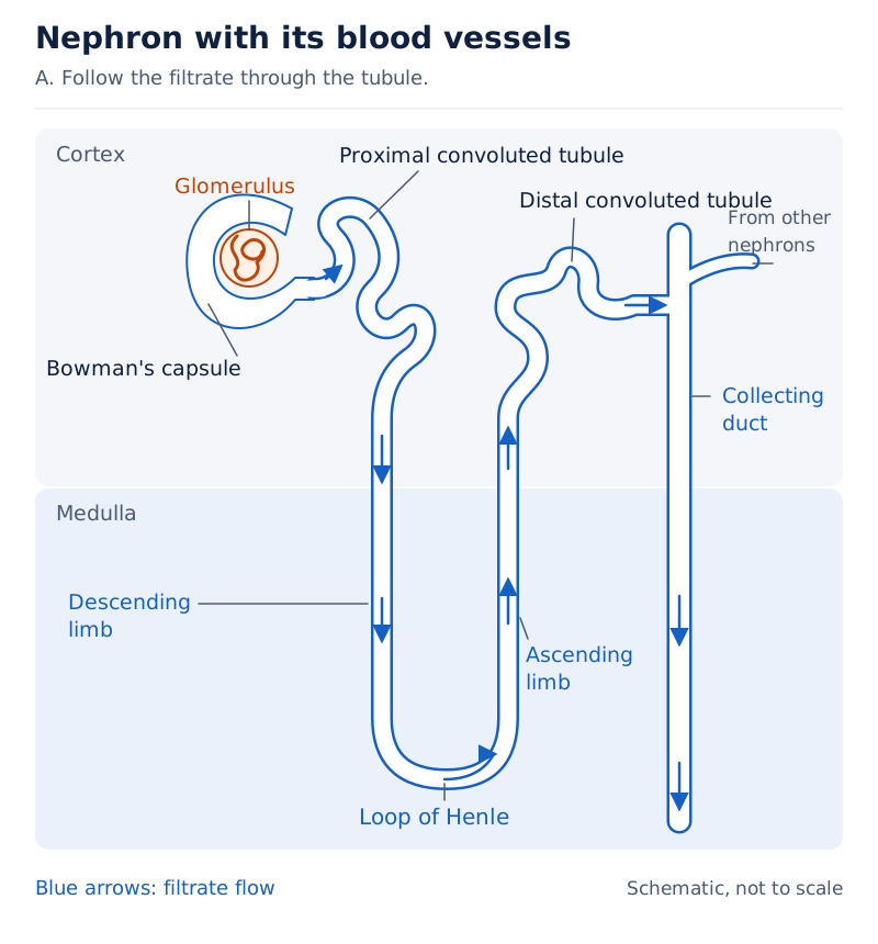
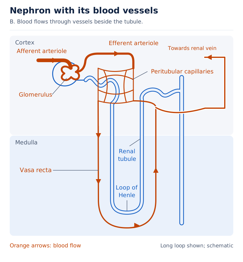
The glomerulus lies within Bowman's capsule near the top. Coiled proximal and distal tubules, the descending and ascending limbs of the loop of Henle, and a collecting duct are labelled. Blood vessels run beside the tubule, including a long loop of small blood vessels labelled vasa recta.
Drawn by One Young India.
The vasa recta are small vessels running parallel to the loop of Henle; they are absent or highly reduced in the majority of nephrons with short loops. The diagram therefore shows two neighbouring systems: a tubule carrying filtrate and vessels carrying blood. Their close arrangement permits exchange without treating the tubule as a blood vessel.
How does blood flow through the kidney's filtering unit?
The renal artery branches within the kidney. An arteriole is a fine arterial branch. The afferent arteriole brings blood into a glomerulus, while the efferent arteriole carries it away. Afferent here means entering; efferent means leaving.
The glomerulus is therefore a capillary tuft positioned between two arterioles. Blood leaves this tuft through a blood vessel, while the liquid filtered from it enters Bowman's capsule. Blood flow and filtrate flow must be traced separately.
What happens beyond the glomerulus?
The efferent arteriole forms a fine network around the renal tubule called the peritubular capillaries. Peritubular means around the tubule. Their position allows substances reabsorbed from the tubular fluid to return to the blood rather than leave in urine.
- The renal artery brings blood to the kidney and divides into smaller branches within the organ.
- An afferent arteriole carries blood into the glomerular capillary tuft enclosed by Bowman's capsule.
- Glomerular blood pressure drives filtration, while the blood remaining in the vessels continues through the tuft.
- The efferent arteriole carries blood away from the glomerulus and forms capillaries around the tubule.
- Following exchange within the kidney, blood is carried away from the organ through the renal vein.
Note: The efferent arteriole is not the ureter. It carries blood away from a glomerulus. The ureter is a separate tube carrying urine away from a kidney towards the urinary bladder.
The surrounding capillaries matter because initial filtration removes useful dissolved materials along with wastes. Returning useful substances requires a route back into circulation. The nephron's tubule and its associated capillaries work together to provide that route.
For labelling, identify where each vessel begins or ends instead of relying on a similar-sounding name. The afferent vessel enters the tuft, the efferent vessel leaves it, and the proximal convoluted tubule continues from the capsule. These three connections carry different fluids at different stages.
How does ultrafiltration produce the initial filtrate?
Ultrafiltration is the fine filtration of blood under pressure through the glomerular filtering membranes. It is also called glomerular filtration. The liquid entering Bowman's capsule is the glomerular filtrate, which will be modified as it passes along the renal tubule.
Blood plasma is the fluid part of blood in which blood cells are suspended. It carries dissolved materials, including nutrients, salts and nitrogenous wastes. Filtration transfers water and many small dissolved substances from plasma into the capsule.
What drives filtration, and what is retained?
Blood pressure in the glomerular capillaries causes filtration. The filtering barrier includes the capillary lining, the lining of Bowman's capsule and a basement membrane, a thin supporting layer between them. The fluid passes through this barrier into the capsule's cavity.
Podocytes are specialised cells of Bowman's capsule arranged with minute spaces between them. These spaces are called filtration slits. Their arrangement forms part of the fine barrier through which the initial filtrate passes.
Glucose is a simple sugar used by cells, and amino acids are the building units of proteins.
Almost all the constituents of plasma except proteins pass into Bowman's capsule. Blood cells remain in the blood rather than entering the normal filtrate. Thus, the initial filtrate contains water, salts, glucose, amino acids and nitrogenous wastes, rather than being whole blood.
The presence of glucose and amino acids in the initial filtrate shows that glomerular filtration does not separate useful substances from wastes according to the body's needs.
What the figure shows
Malpighian body or renal corpuscle
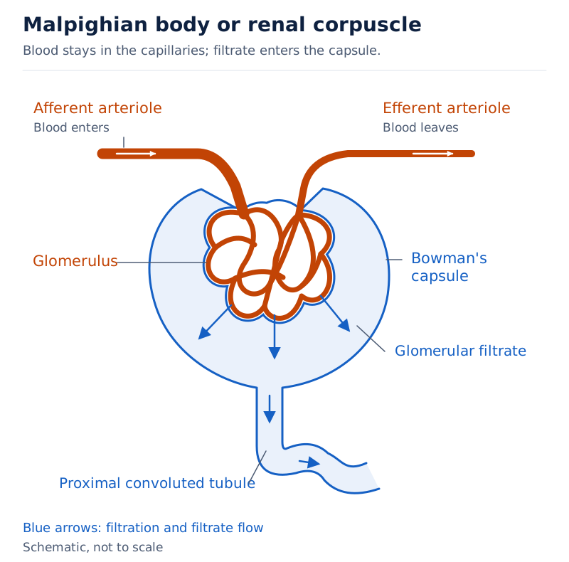
A red branching capillary tuft lies within a blue cup-shaped Bowman's capsule. The afferent arteriole enters above, and the efferent arteriole leaves above. The capsule continues below into the labelled proximal convoluted tubule.
Drawn by One Young India.
In a labelled drawing, keep the glomerular blood vessels distinct from the space receiving filtrate. The vessel leaving the tuft must remain connected with the blood pathway. The capsule's continuation must connect with the tubule, which receives the filtered liquid.
The filtrate is not yet final urine. If the filtering step were the whole process, useful nutrients and a large amount of water would be lost. Further processing by the renal tubule is therefore central to understanding urine formation.
Note: Filtration is pressure-driven and does not select substances according to whether they are useful. Selective recovery of useful substances occurs after filtration, as fluid passes along the tubule.
How does selective reabsorption conserve useful substances?
Selective reabsorption is the return of substances from the tubular filtrate to the blood according to the body's requirements. Glucose, amino acids, salts and a major amount of water are reabsorbed as fluid passes through the nephron.
The word reabsorption refers to taking substances back from the filtrate. It does not mean bringing fresh blood into the glomerulus. The substances have already left the blood during filtration and are now recovered through the tubular wall.
Where does reabsorption occur?
Electrolytes are substances that provide ions when dissolved in water. The proximal convoluted tubule is a major site. Its lining has a brush border, a surface with tiny projections that increases the area available for reabsorption. Nearly all essential nutrients and 70 to 80 per cent of water and electrolytes are reabsorbed by this segment.
Reabsorbing appropriate amounts of electrolytes helps maintain the composition of body fluids. The same general principle applies to conserving water rather than allowing the entire filtered quantity to be lost.
Some substances, including glucose, amino acids and sodium ions, are reabsorbed by active transport, movement requiring cellular energy. Water reabsorption in the initial segments occurs passively, without being directly driven by cellular energy expenditure.
Permeable means allowing passage; impermeable means not allowing passage. The descending limb of the loop of Henle is permeable to water but almost impermeable to electrolytes. The ascending limb is impermeable to water but allows electrolyte transport. These differences help alter the concentration of the tubular fluid.
Further reabsorption of sodium ions and water occurs conditionally in the distal convoluted tubule. Large amounts of water could also be reabsorbed from the collecting duct to produce concentrated urine. Reabsorption therefore continues beyond the proximal tubule.
Why does the quantity of urine vary?
The amount of water reabsorbed depends on the excess water present in the body and on the dissolved waste that must be excreted. Urine formation combines waste removal with osmoregulation, the regulation of body water and dissolved salts.
A comparison of about 180 litres of daily filtrate with about 1.5 litres of urine shows the scale of recovery. Nearly 99 per cent of the filtrate is reabsorbed. The large initial volume therefore does not imply an equally large daily loss of water.
This comparison concerns filtrate and urine, not two measurements of the blood itself. It also explains why a urine sample has a different composition from the initial filtrate. Reabsorption changes both the amount of fluid and the substances remaining in it.
How does tubular secretion complete the processing of filtrate?
Tubular secretion is the addition of certain substances to the filtrate by cells of the renal tubule. It supplements filtration by transferring substances into the tubular fluid during urine formation. Its direction is opposite to the recovery of substances during reabsorption.
Tubular cells secrete hydrogen ions, potassium ions and ammonia into the filtrate. These processes help maintain ionic balance, the appropriate proportions of dissolved ions, and acid-base balance, the regulation of acidity in body fluids.
How are the three processes different?
Filtration begins urine formation at the glomerulus. Reabsorption takes substances out of the filtrate and returns them to the blood. Secretion adds particular substances to the filtrate. Together, the three processes account for the difference between blood plasma and the urine eventually released.
| Feature | Ultrafiltration | Selective reabsorption | Tubular secretion |
|---|---|---|---|
| General location | Glomerulus and Bowman's capsule | Along the renal tubule | Through tubular cells |
| Direction relative to tubular fluid | Forms the initial filtrate | Removes substances from filtrate | Adds substances to filtrate |
| Relationship to blood | Filters fluid from glomerular blood | Returns reabsorbed substances to blood | Helps adjust the ionic and acid-base composition of body fluids |
| Examples | Water and small dissolved substances enter the capsule | Glucose, amino acids, salts and much water are recovered | Hydrogen ions, potassium ions and ammonia enter the filtrate |
| Main significance | Produces fluid for further processing | Conserves useful substances and water | Contributes to ionic and acid-base balance |
The proximal convoluted tubule can secrete hydrogen ions and ammonia. The distal convoluted tubule can secrete hydrogen ions, potassium ions and ammonia. The collecting duct also participates in selective secretion of hydrogen and potassium ions.
A single tubular region can therefore participate in both reabsorption and secretion. These terms describe the direction and role of movement, rather than naming mutually exclusive kinds of tubule. The presence of reabsorption does not mean that secretion is absent.
Note: Tubular secretion and the release of urine are different processes. Secretion changes the composition of fluid inside the kidney. Urination releases urine already formed and stored in the urinary bladder.
How do plasma, filtrate and urine differ, and how is urine released?
Blood plasma, glomerular filtrate and urine represent different stages of kidney processing. Plasma arrives as part of blood, filtrate enters Bowman's capsule, and final urine remains after tubular reabsorption and secretion. They should not be described as identical fluids.
| Feature | Blood plasma | Initial filtrate | Normal urine |
|---|---|---|---|
| Location in this pathway | Within the renal blood vessels | Within Bowman's capsule and the beginning of the tubule | Leaves the kidney through the ureter |
| Water | Liquid medium carrying dissolved substances | Passes through the filtering barrier | Remains after extensive water reabsorption |
| Proteins | Present in plasma | Retained by the normal filtering barrier | Not normal constituents in appreciable amounts |
| Glucose | Transported in the blood | Enters during filtration | Normally absent after reabsorption |
| Nitrogenous wastes | Carried towards the kidneys | Enter the fluid being processed | Eliminated from the body, including urea |
| Salts | Present in dissolved form | Enter during filtration | Present after tubular adjustment of ions |
Normal urine is a light-yellow watery fluid with a characteristic odour. It is slightly acidic, with a typical pH of about 6. Here, pH is a measure of acidity or alkalinity. Urine characteristics can change under different conditions.
An adult human excretes, on average, 1 to 1.5 litres of urine per day and, on average, 25 to 30 grams of urea per day. These are average values, not fixed quantities produced by every individual under every condition.
What is the route from the collecting ducts to the exterior?
The urethral sphincter is a muscle controlling the bladder outlet. It relaxes as the bladder wall contracts during the release of urine.
- Collecting ducts receive fluid from nephrons and convey it towards the renal pelvis through the medullary pyramids and calyces.
- Urine passes from the renal pelvis into the ureter associated with that kidney.
- The ureter carries urine to the urinary bladder, where urine is stored.
- Filling stretches the bladder wall and produces signals involved in the urge to urinate.
- Contraction of the bladder wall, together with relaxation of the urethral sphincter, releases urine through the urethra.
The release of urine is called micturition. Bladder activity is under nervous control, and we can usually control the urge to urinate. Storage and release occur after the kidney has formed urine.
In a complete explanation, connect each stage with the next: blood is filtered, useful materials are recovered, additional substances are secreted, and urine is transported and stored. The bladder stores the final product; it does not perform the kidney's filtration and reabsorption.
Glossary
- Excretion — The biological process that removes harmful metabolic waste substances from the body.
- Renal pelvis — The funnel-shaped space inside a kidney that receives urine before it enters the ureter.
- Nephron — A functional unit of the kidney containing a glomerulus and a renal tubule.
- Glomerulus — A tuft of blood capillaries within Bowman's capsule where initial filtration occurs.
- Bowman's capsule — The double-walled, cup-like beginning of the renal tubule that surrounds the glomerulus and receives filtrate.
- Malpighian body — The renal corpuscle formed by the glomerulus together with its surrounding Bowman's capsule.
- Afferent arteriole — The fine arterial vessel that carries blood into the glomerular capillary tuft.
- Efferent arteriole — The vessel carrying blood away from the glomerulus and forming capillaries around the tubule.
- Ultrafiltration — Fine, pressure-driven filtration through the glomerular barrier into the cavity of Bowman's capsule.
- Glomerular filtrate — Fluid formed from blood plasma by filtration and received within Bowman's capsule.
- Selective reabsorption — Recovery of substances from tubular filtrate into the blood according to the body's requirements.
- Tubular secretion — Addition of substances to the filtrate by tubular cells during the formation of urine.
- Collecting duct — A straight tube receiving fluid from several nephrons and conducting it towards the renal pelvis.
- Osmoregulation — Regulation of body water and dissolved salts to maintain an appropriate internal fluid composition.
- Micturition — The release of stored urine from the urinary bladder through the urethra.
Common errors and misconceptions
- Misconception: Excretion and egestion mean the same thing. Correct: Excretion removes metabolic wastes; egestion removes undigested food from the digestive tract.
- Misconception: Urea is made in the kidneys. Correct: The liver converts ammonia into urea; blood carries urea to the kidneys for removal.
- Misconception: The ureter carries urine out of the body. Correct: A ureter connects a kidney with the bladder; the urethra carries urine from the bladder to the exterior.
- Misconception: The efferent arteriole carries urine away from a nephron. Correct: It carries blood away from the glomerulus; filtrate continues through the renal tubule.
- Misconception: Filtration removes only waste substances. Correct: Useful substances, including glucose and amino acids, also enter the initial filtrate and are subsequently reabsorbed.
- Misconception: Reabsorption adds wastes to the filtrate. Correct: Reabsorption returns substances to the blood; tubular secretion adds certain substances to the filtrate.
- Misconception: All filtered water leaves as urine. Correct: A major amount of water is reabsorbed, and the amount recovered depends on the body's water and waste-removal requirements.
- Misconception: Urine is formed in the bladder. Correct: Kidneys form urine; the bladder stores it until micturition releases it through the urethra.
Exam-style questions with model answers
Q1. Distinguish between a ureter and the urethra by stating the route followed by urine in each. [2 marks]
- A ureter carries urine from a kidney to the urinary bladder.
- The urethra carries stored urine from the urinary bladder to the outside of the body.
Q2. Name the organ that converts ammonia into urea and explain how urea reaches the organs that remove it. [2 marks]
- The liver converts ammonia produced during metabolism into the less toxic nitrogenous waste urea.
- Urea enters the blood, which carries it to the kidneys for removal in urine.
Q3. State one excretory contribution each of the lungs, liver and sweat glands. [3 marks]
- The lungs remove carbon dioxide and significant quantities of water from the body, contributing to the elimination of gaseous wastes.
- The liver secretes bile containing substances such as bile pigments and cholesterol; most of these ultimately leave with digestive wastes.
- Sweat glands release a watery secretion containing sodium chloride, small amounts of urea and lactic acid, although sweating primarily cools the body.
Q4. Describe the kidney's cortex, medullary pyramids, renal columns and renal pelvis, giving one structural point for each. [4 marks]
- The cortex forms the outer internal zone of the kidney, lying beneath the tough renal capsule that covers the organ.
- The inner medulla is divided into conical masses called medullary pyramids, which project into the extensions of the renal pelvis.
- Renal columns are extensions of the cortex between adjacent medullary pyramids; they are also called columns of Bertini.
- The renal pelvis is a broad funnel-shaped space internal to the hilum, with extensions called calyces, leading towards the ureter.
Q5. Explain ultrafiltration at the Malpighian body. Include its location, driving force, barrier, substances entering the filtrate and substances retained in blood. [5 marks]
- Ultrafiltration occurs at the renal corpuscle, where the glomerular capillary tuft is enclosed by Bowman's capsule at the beginning of the nephron.
- Blood pressure within the glomerular capillaries provides the force that drives fluid through the filtering barrier into the capsule.
- The barrier includes the capillary lining, the lining of Bowman's capsule and the basement membrane between them; podocytes leave fine filtration slits.
- Water and small dissolved substances enter the filtrate, including glucose, amino acids, salts and nitrogenous wastes that will undergo further tubular processing.
- Plasma proteins and blood cells are retained in the blood under normal conditions, so the liquid entering Bowman's capsule is not whole blood.
Q6. Trace fluid from Bowman's capsule to its release outside the body in six stages. Include tubular processing, collection, transport, storage and release. [6 marks]
- Filtrate in Bowman's capsule enters the proximal convoluted tubule, where extensive reabsorption recovers useful nutrients and much water from the tubular fluid.
- It continues through the descending and ascending limbs of the loop of Henle, whose different permeabilities help alter the fluid's concentration.
- It reaches the distal convoluted tubule and then a collecting duct, where further reabsorption and selective secretion adjust its composition.
- The collecting ducts convey the fluid towards the renal pelvis through the medullary pyramids and calyces; urine then enters the ureter.
- The ureter carries urine from the kidney to the urinary bladder, which stores it until the process of release occurs.
- Bladder contraction and relaxation of the urethral sphincter release urine through the urethra to the exterior, a process called micturition.
Q7. Glucose is present in an initial filtrate sample but absent from the final urine of a healthy person. Explain this in three points, naming the processes involved. [3 marks]
- Glomerular filtration allows glucose dissolved in blood plasma to enter Bowman's capsule along with water and other small dissolved substances.
- Selective reabsorption subsequently returns glucose from the tubular filtrate to the blood; the proximal convoluted tubule is a major site of nutrient recovery.
- Glucose reabsorption uses active transport, and essentially all filtered glucose is recovered under normal conditions, so glucose is normally absent from final urine.
Q8. Distinguish selective reabsorption from tubular secretion by direction and function. Give four separate points. [4 marks]
- Selective reabsorption takes substances out of the tubular filtrate and returns them to the blood through the renal tubular wall.
- It conserves useful materials such as glucose, amino acids, salts and much water, helping maintain the body's fluid composition.
- Tubular secretion adds particular substances to the filtrate through the activity of tubular cells; examples include hydrogen ions, potassium ions and ammonia.
- Secretion contributes to ionic and acid-base balance, while the secreted substances become part of the fluid being processed into urine.
Key takeaways
- The kidneys remove nitrogenous wastes, while the lungs, liver and skin also contribute to the elimination of excretory substances.
- The urinary system contains two kidneys, two ureters, a urinary bladder and a urethra, with different roles in urine handling.
- A kidney has an outer cortex and inner medulla; medullary pyramids project into calyces associated with the renal pelvis.
- The Malpighian body consists of a glomerulus enclosed by Bowman's capsule at the beginning of the renal tubule.
- Ultrafiltration forms an initial filtrate containing useful dissolved substances as well as wastes; it does not produce final urine immediately.
- Selective reabsorption recovers useful substances and much water, while tubular secretion adds certain substances to the filtrate.
- The afferent and efferent arterioles carry blood; the renal tubule carries filtrate, and the ureter carries urine.
- Urine is formed in the kidneys, stored in the bladder and released through the urethra during micturition.
Test yourself
What two structures together form a Malpighian body?
The glomerular capillary tuft and its surrounding Bowman's capsule together form the Malpighian body or renal corpuscle.
Which arteriole enters a glomerulus, and which leaves it?
The afferent arteriole brings blood into the glomerulus, and the efferent arteriole carries blood away from it.
Why is glomerular filtrate not identical to final urine?
Useful substances and much water are reabsorbed, while tubular cells add certain substances by secretion as the filtrate moves through the nephron.
What is the main force driving ultrafiltration?
Blood pressure within the glomerular capillaries drives filtration through the barrier into Bowman's capsule.
What is the difference between the renal capsule and Bowman's capsule?
The renal capsule covers the whole kidney. Bowman's capsule is the cup-like beginning of a renal tubule enclosing one glomerulus.
How does the brush border assist the proximal convoluted tubule?
Its tiny surface projections increase the area available for reabsorption from the tubular filtrate.
What determines how much water is reabsorbed?
Water reabsorption depends on the excess water in the body and the amount of dissolved waste that must be excreted.
Why does urine in the bladder not indicate urine formation there?
The kidneys form urine, which the ureters deliver to the bladder. The bladder stores it until release through the urethra.
Try it
ICSE Class 10 Biology: The Human Excretory System and Osmoregulation
Design a 2-step scenario interactive for a study note.
1What is the primary function of excretion in the human body?
Excretion is the process of eliminating toxic end-products of metabolism, excess mineral salts, and water, not undigested food residue, which is egestion.
Excretion is the essential physiological process by which the human body eliminates toxic end-products of metabolism, excess mineral salts, and water to maintain internal homeostasis.
Bile pigments are a type of excretory substance, but they are not the primary function of excretion.
2What is the main difference between the renal cortex and the renal medulla?
The renal cortex is the outer, dark-red, granular zone, while the renal medulla is the inner, paler, striated zone.
The renal cortex contains the Malpighian bodies, proximal convoluted tubules, and distal convoluted tubules, while the renal medulla is organized into conical masses termed renal pyramids with parallel loops of Henle and collecting ducts.
The renal cortex and renal medulla have different functions, but they are not responsible for filtering waste and storing urine, respectively.
3What is the function of the Loop of Henle in the nephron?
The Loop of Henle is involved in water and electrolyte transport, not glucose and amino acid reabsorption.
The Distal Convoluted Tubule (DCT) is responsible for regulated salt reabsorption, proton excretion, and potassium secretion, not the Loop of Henle.
The Loop of Henle features a thin descending limb (permeable to water, impermeable to electrolytes) and a thicker ascending limb (impermeable to water, actively transporting sodium and chloride ions into the medullary interstitium).
The human excretory system is a complex process that involves the elimination of toxic substances, excess mineral salts, and water to maintain internal homeostasis. The nephron is the functional unit of the kidney, responsible for filtering waste and excess substances from the blood. Understanding the structure and function of the nephron is crucial for understanding the human excretory system.