
Molecular basis of Inheritance | ISC Class 12 Biology Notes
On this page
This note covers deoxyribonucleic acid (DNA), ribonucleic acid (RNA), genetic material, chromosome packaging, replication, transcription, the genetic code, protein synthesis, regulation of gene expression, the Human Genome Project and DNA fingerprinting.
How does the structure of DNA explain inheritance?
What joins the building blocks?
Nucleic acids are long molecules built from repeating units called nucleotides. A nucleotide contains a nitrogenous base, a pentose or five-carbon sugar, and a phosphate group. DNA contains deoxyribose sugar; RNA contains ribose. A base joined to sugar alone forms a nucleoside.
The bases adenine (A) and guanine (G) are purines; cytosine (C), thymine (T) and uracil (U) are pyrimidines. These are the two structural classes of nitrogenous bases. DNA contains A, G, C and T; RNA contains U in place of T.
The symbols 5′ and 3′ refer to the fifth and third carbon positions of the sugar. A 3′-5′ phosphodiester linkage joins successive nucleotides through phosphate. The resulting polynucleotide, a chain of many nucleotides, has a sugar-phosphate backbone with bases projecting from it.
The 5′ end has a free phosphate group, whereas the 3′ end has a free hydroxyl group, written OH. A base attaches at the sugar's 1′ carbon through an N-glycosidic linkage, a bond involving nitrogen in the base.
What did the double-helix model establish?
Friedrich Miescher identified the nuclear substance called nuclein in 1869. In 1953, James Watson and Francis Crick proposed the double-helix model using X-ray diffraction data produced by Maurice Wilkins and Rosalind Franklin. Erwin Chargaff's observations supported equal amounts of A and T, and of G and C, in double-stranded DNA.
- The two chains are antiparallel, meaning they run in opposite directions: one 5′ to 3′ and the other 3′ to 5′.
- A pairs with T through two hydrogen bonds; G pairs with C through three. A paired set is a base pair, abbreviated bp.
- A purine pairs with a pyrimidine, giving an approximately uniform separation between the chains.
- The chains form a right-handed helix with a pitch, or distance per complete turn, of 3.4 nanometres. A nanometre, abbreviated nm, is one billionth of a metre.
- There are roughly 10 bp per turn, with approximately 0.34 nm between consecutive base pairs. Stacking of base pairs and hydrogen bonding stabilise the helix.
Complementarity means the bases of one strand specify those of its partner. Each strand can template a new complementary strand.
What the figure shows
DNA double helix
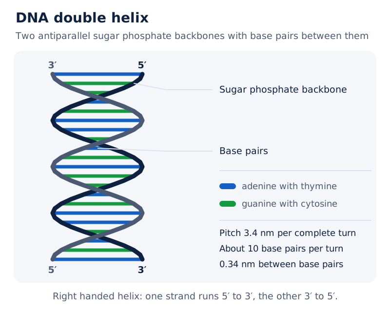
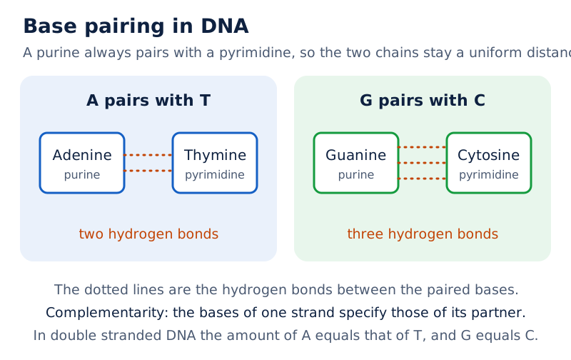
The drawing shows two winding sugar-phosphate backbones surrounding paired bases. Labels identify the backbone and base pairs; separate coloured pairs identify adenine with thymine and guanine with cytosine.
Drawn by One Young India.
How is DNA packaged into chromosomes?
Prokaryotes are organisms whose cells lack a defined nucleus; eukaryotes have a defined nucleus. A typical mammalian cell contains approximately 2.2 metres of DNA, much longer than the dimensions of its nucleus.
In the bacterium Escherichia coli, abbreviated E. coli, negatively charged DNA associates with positively charged proteins in a region called the nucleoid. Its DNA forms large loops held by proteins rather than lying scattered throughout the cell.
How do nucleosomes form?
Eukaryotic DNA associates with histones, positively charged basic proteins. Proteins are molecules made from amino acids, their building units. Histones contain abundant lysine and arginine, amino acids whose side chains carry positive charges.
- Eight histone molecules form a histone octamer, an eight-part protein core.
- Negatively charged DNA wraps around this positively charged core to form a nucleosome. A typical nucleosome contains 200 bp of DNA helix.
- Nucleosomes repeat along chromatin, the thread-like material of the nucleus, producing a beads-on-string appearance under an electron microscope.
- The nucleosome chain is packaged into chromatin fibres, which coil and condense into chromosomes during metaphase, the cell-division stage when chromosomes are highly condensed.
Higher levels of packaging require additional non-histone chromosomal proteins, chromosome-associated proteins other than histones.
Euchromatin is loosely packed, stains lightly and is said to be transcriptionally active, meaning its DNA can be copied into RNA. Heterochromatin is densely packed, stains darkly and is transcriptionally inactive.
What the figure shows
Nucleosome and beads-on-string chromatin
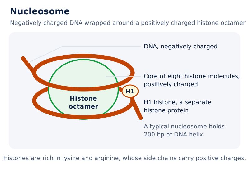
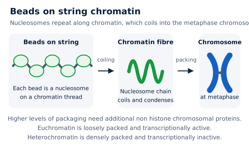
The nucleosome drawing labels DNA wrapped around a histone octamer, the histone core and H1 histone, a separately labelled histone protein. The accompanying electron micrograph shows bead-like structures along chromatin threads.
Drawn by One Young India.
Which experiments identified DNA as genetic material?
What did Griffith demonstrate?
Genetic material carries inherited information. In 1928, Frederick Griffith studied Streptococcus pneumoniae, a bacterium that causes pneumonia. The smooth, or S, strain had a polysaccharide coat, a covering made of linked sugar units. It was virulent, meaning disease-causing. The rough, or R, strain lacked this coat and did not produce pneumonia in the mice.
| Material injected into mice | Observation | Meaning |
|---|---|---|
| Living S bacteria | Mice died | The smooth strain was virulent |
| Living R bacteria | Mice survived | The rough strain did not cause pneumonia |
| Heat-killed S bacteria | Mice survived | Killed S cells did not cause the disease |
| Heat-killed S bacteria mixed with living R bacteria | Mice died; living S bacteria were recovered | Material from dead S cells transformed living R cells |
Transformation was the inherited change that enabled R bacteria to form a smooth coat and become virulent. Griffith inferred a transforming principle, the material responsible for this change, but did not identify its biochemical nature.
How was the transforming principle identified?
Oswald Avery, Colin MacLeod and Maclyn McCarty purified substances from heat-killed S bacteria. DNA alone caused transformation of living R bacteria into S bacteria. Enzymes are biological catalysts, substances that speed reactions. Proteases digest proteins and RNases digest RNA; neither stopped transformation.
DNase, an enzyme that digests DNA, inhibited transformation. This result identified DNA as the hereditary material responsible for the change.
What did Hershey and Chase trace?
Alfred Hershey and Martha Chase used bacteriophages, viruses that infect bacteria, in 1952. They tested whether viral DNA or protein entered E. coli. Radioactive phosphorus labelled the phage DNA; radioactive sulphur labelled the phage protein coat.
- Prepare phages with radioactively labelled DNA and a separate set with labelled protein coats.
- Allow the labelled phages to attach to bacteria and begin infection.
- Agitate the mixture in a blender to detach viral coats from the bacteria.
- Use centrifugation, separation by rapid spinning, to separate bacterial cells from detached viral material.
- Check the bacteria for radioactivity. It entered with labelled DNA, whereas the bacteria exposed to protein-labelled phages were not radioactive.
DNA entered the bacteria and directed production of new phages.
Why is DNA a more stable genetic material than RNA?
Genetic material must replicate, remain chemically and structurally stable, allow slow heritable changes and express inherited characters. A mutation is a change in genetic material. Stability preserves information, while the possibility of mutation allows variation needed for evolution.
How do the two nucleic acids compare?
| Feature | DNA | RNA |
|---|---|---|
| Sugar | Deoxyribose | Ribose |
| Characteristic pyrimidine | Thymine | Uracil in place of thymine |
| Hydroxyl group at the 2′ position | Lacks the additional hydroxyl group present in ribose | Has an additional reactive hydroxyl group |
| Chemical behaviour | Less reactive and structurally more stable | More reactive and easily degraded |
| Genetic role | Genetic material in most organisms | Genetic material in some viruses |
| Protein synthesis | Depends on RNA for protein synthesis | Can directly code for protein synthesis |
Complementary base pairing allows both DNA and RNA to direct duplication. Both can mutate, but unstable RNA mutates at a faster rate. DNA's greater stability makes it better suited to storing genetic information. Thymine also contributes additional stability compared with uracil.
RNA mostly functions as a messenger and also performs adapter, structural and, in some cases, catalytic roles. Catalytic RNA is called a ribozyme. RNA's functions help explain why protein synthesis is organised around RNA.
What is the central dogma?
The central dogma, proposed by Francis Crick, describes information flow from DNA to RNA to protein. Transcription copies DNA information into RNA. Translation uses RNA information to assemble a chain of amino acids, called a polypeptide.
Reverse transcription is information transfer from RNA to DNA, found in some viruses. The RNA-world idea places RNA before DNA as genetic material. Evidence suggests that essential processes, including translation and RNA processing, evolved around RNA; chemical modifications made DNA more stable.
How does DNA replicate, and what proves that it is semiconservative?
Replication is the copying of DNA. In semiconservative replication, the two parental strands separate, each directs formation of a complementary strand, and each daughter DNA molecule contains one parental strand and one newly synthesised strand.
What happens at a replication fork?
A replication fork is the small opened region of the DNA helix where copying occurs. DNA-dependent DNA polymerase is the enzyme that uses a DNA template to join deoxyribonucleotides into a new DNA strand.
- Replication begins at a defined DNA region called the origin of replication. DNA polymerase cannot initiate replication on its own.
- The helix opens locally, exposing parental strands that provide complementary templates.
- DNA polymerase builds new DNA in the 5′ to 3′ direction. Deoxyribonucleoside triphosphates supply both building units and energy.
- Copying is continuous on the template running 3′ to 5′, but discontinuous on the template running 5′ to 3′.
- DNA ligase, the enzyme that joins DNA fragments, connects the discontinuously synthesised pieces.
Both new strands grow 5′ to 3′. In eukaryotes, replication occurs during the S phase, the DNA-synthesis phase of the cell cycle.
How did Meselson and Stahl test the model?
In 1958, Matthew Meselson and Franklin Stahl grew E. coli for many generations with ¹⁵N, the heavy, non-radioactive isotope of nitrogen. An isotope is a form of an element with a different atomic mass. They then transferred the cells to medium containing normal ¹⁴N.
Extracted double-stranded DNA was separated in a caesium chloride density gradient, a solution with a range of densities. DNA containing heavy nitrogen could thus be distinguished from light DNA and intermediate-density DNA.
| Sample | DNA observed | Interpretation |
|---|---|---|
| Before transfer | Heavy DNA | Both strands contained heavy nitrogen |
| After one generation, 20 minutes | Hybrid DNA of intermediate density | Each molecule contained a heavy parental strand and a light new strand |
| After two generations, 40 minutes | Equal amounts of hybrid and light DNA | Replication produced both heavy-light and light-light molecules |
Here hybrid DNA means a double helix with one heavy and one light strand. The generation time applies to these experimental conditions.
Taylor and colleagues used radioactive thymidine, a labelled DNA component, to detect newly synthesised DNA in chromosomes of Vicia faba, the faba bean, in 1958. Their experiments demonstrated semiconservative replication in chromosomes as well.
What the figure shows
Meselson and Stahl's experiment
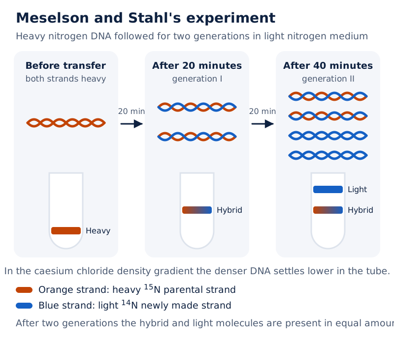
The drawing follows heavy DNA through two generations. Coloured strands distinguish old and new DNA, while centrifuge tubes show a heavy band initially, a hybrid band after generation I, and light and hybrid bands after generation II.
Drawn by One Young India.
How is a transcription unit organised?
Transcription copies information from one DNA strand into RNA. Unlike replication of the full DNA, transcription copies a segment. Complementary pairing is retained, but RNA uses uracil opposite adenine. Only one strand is copied per transcription unit.
What determines the start, template and end?
A transcription unit comprises a promoter, a structural gene and a terminator. The promoter is the DNA sequence where RNA polymerase binds. The structural gene is the region transcribed. The terminator usually defines where transcription ends.
DNA-dependent RNA polymerase makes RNA using DNA as its template. It builds RNA 5′ to 3′ while using the template strand running 3′ to 5′. The opposite coding strand has the RNA sequence except for T in place of U.
Positions are described relative to the coding strand. Upstream means towards its 5′ end, where the promoter lies; downstream means towards its 3′ end, where the terminator lies. Promoter position defines the template strand.
How do bacteria carry out transcription?
- Initiation: RNA polymerase associates with the promoter. The initiation factor sigma, represented by the Greek symbol σ, assists the start.
- Elongation: RNA polymerase joins ribonucleotides using nucleoside triphosphates, the three-phosphate forms of nucleosides, as substrates.
- Termination: at the terminator region, the newly formed RNA and polymerase separate from the DNA. The termination factor rho, represented by ρ, assists termination.
- The bacterial RNA can become available for translation. Many times translation begins before the complete messenger RNA has been transcribed because the processes share the same compartment.
If complementary RNA copies were made from both DNA strands, they would have different sequences and could pair with each other. Double-stranded RNA formed in this way would prevent translation. Complementary sequences must therefore not be mistaken for identical sequences.
What are the types of RNA, and how is eukaryotic RNA processed?
Which RNA performs each function?
| RNA type | Full name | Role |
|---|---|---|
| mRNA | Messenger RNA | Provides the template for protein synthesis |
| tRNA | Transfer RNA | Reads the genetic code and brings the corresponding amino acid |
| rRNA | Ribosomal RNA | Provides structural and catalytic functions in ribosomes, the sites of protein synthesis |
| hnRNA | Heterogeneous nuclear RNA | Forms the primary precursor of messenger RNA in eukaryotes |
| snRNA | Small nuclear RNA | Participates in removal of introns during RNA splicing |
A gene is the functional unit of inheritance. It need not code for a protein: DNA sequences specifying tRNA and rRNA are also genes. A cistron is a DNA segment coding for a polypeptide.
A monocistronic transcription unit contains one cistron and is found mostly in eukaryotes. A polycistronic transcription unit contains several cistrons and is found mostly in bacteria or other prokaryotes. Recombination is the exchange of genetic material. A recon is the smallest genetic unit capable of recombination; a muton is the smallest genetic unit capable of mutation.
What changes turn hnRNA into mature mRNA?
Eukaryotic nuclear RNA polymerases have different roles. RNA polymerase I transcribes major ribosomal RNAs. RNA polymerase II transcribes hnRNA. RNA polymerase III transcribes tRNA, another ribosomal RNA and small nuclear RNAs.
The initial RNA transcript contains exons, sequences retained in mature RNA, and introns, intervening sequences removed during processing. Processing produces functional messenger RNA.
- Splicing: introns are removed and exons are joined in a defined order.
- Capping: an unusual nucleotide, methyl guanosine triphosphate, is added to the 5′ end.
- Tailing: 200 to 300 adenylate residues, adenine-containing nucleotide units, are added at the 3′ end without a DNA template.
Fully processed mRNA leaves the nucleus for translation.
What the figure shows
RNA processing in eukaryotes
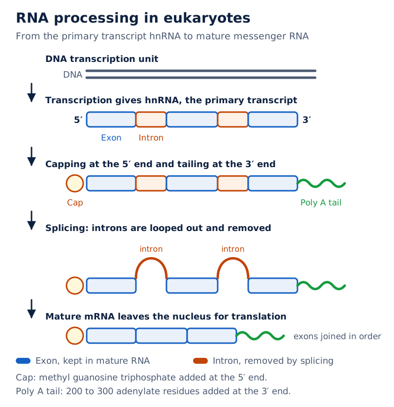
The drawing shows RNA emerging from DNA, a cap at the 5′ end, labelled intron and exon regions, and a poly-A tail, a chain of adenylate residues, at the 3′ end. Looped segments illustrate intron removal before the final messenger RNA.
Drawn by One Young India.
How does the genetic code specify amino acids?
The genetic code connects the order of bases in mRNA with the order of amino acids in a polypeptide. A codon is a sequence of three successive mRNA nucleotides specifying an amino acid or a stop signal.
How was the code deciphered?
George Gamow proposed a three-base code. Four available bases in each of three positions give 4 × 4 × 4 = 64 combinations, sufficient to specify 20 amino acids.
Har Gobind Khorana developed chemical methods for making RNA with defined base combinations. Marshall Nirenberg's cell-free protein-synthesis system helped decipher the code. Severo Ochoa's enzyme, polynucleotide phosphorylase, helped synthesise RNA in a template-independent manner.
Which features must be distinguished?
- Triplet: each codon contains three nucleotides; 61 codons specify amino acids and three serve as stop signals.
- Degenerate: some amino acids are specified by more than one codon. This does not mean one codon normally specifies several amino acids.
- Contiguous reading: codons are read successively without punctuation between them.
- Nearly universal: a codon generally has the same meaning across organisms. Exceptions occur in mitochondria, energy-releasing cell structures, and some protozoans, single-celled eukaryotic organisms.
- Start signal: AUG specifies methionine, an amino acid, and also initiates translation.
- Stop signals: UAA, UAG and UGA do not specify amino acids and terminate translation.
For example, UUU and UUC both specify the amino acid phenylalanine. Thus a known mRNA sequence specifies its amino-acid sequence, but an amino-acid sequence may not uniquely reveal the original RNA sequence because of degeneracy.
A reading frame is the grouping of successive bases into triplets. Inserting or deleting one or two bases shifts the frame from that point. Inserting or deleting three bases, or a multiple of three, leaves the downstream frame unaltered while changing the number of codons.
A point mutation, a change involving a single base pair, can alter a protein. In sickle-cell anaemia, one such change in the beta-globin gene replaces the amino acid glutamate with valine. Beta-globin is a component of haemoglobin, the oxygen-carrying blood protein.
How does translation assemble a polypeptide?
Translation joins amino acids in the sequence specified by mRNA. The links between successive amino acids are peptide bonds. The process requires mRNA as a template, tRNA as an adapter, and ribosomes containing rRNA and proteins.
Why is tRNA an adapter?
A tRNA has an anticodon, a triplet complementary to an mRNA codon, and an amino-acid acceptor end, linking codon recognition to amino-acid delivery. Its secondary structure resembles a clover leaf; its compact shape resembles an inverted L.
Adenosine triphosphate, abbreviated ATP, supplies energy for activating amino acids. An amino acid is attached to its corresponding tRNA in charging, also called aminoacylation.
What are the stages of protein synthesis?
- Charging: amino acids are activated using ATP and linked to their corresponding tRNAs.
- Initiation: a ribosome binds mRNA at AUG, the start codon, recognised by an initiator tRNA, a specialised tRNA used at the start.
- Codon recognition: charged tRNAs successively pair their anticodons with complementary codons in mRNA.
- Elongation: peptide bonds form between amino acids, and the ribosome moves from codon to codon as the polypeptide grows.
- Termination: a release factor, a protein that ends translation, recognises a stop codon and releases the completed polypeptide.
Ribosomes have a small and a large subunit. The large subunit brings amino acids close enough for bond formation. Bacterial ribosomal RNA catalyses peptide-bond formation, demonstrating that a catalyst need not be a protein.
A translational unit is the mRNA sequence bounded by a start codon and a stop codon that specifies a polypeptide. Untranslated regions, abbreviated UTRs, lie before the start codon and after the stop codon and support efficient translation.
Note: Stop codons have no corresponding tRNAs. Termination involves a release factor, whereas addition of an amino acid involves a charged tRNA. A stop signal is therefore not an extra amino acid in the finished chain.
How does the lac operon regulate gene expression?
Gene expression is the use of genetic information to produce a functional product. In prokaryotes, control of transcriptional initiation is the predominant point of regulation.
An operon is a group of structural genes regulated together through a common promoter and associated control sequences. François Jacob and Jacques Monod explained the lac operon in E. coli; lac refers to lactose, a sugar made of glucose and galactose units.
What do its genes and control regions do?
| Symbol or component | Meaning | Function |
|---|---|---|
| i | Regulatory gene; i derives from inhibitor | Produces the repressor, a protein that blocks transcription |
| p | Promoter | Provides the RNA-polymerase binding site |
| o | Operator, the regulatory DNA region | Binds the repressor |
| z | Structural gene for beta-galactosidase | Produces an enzyme that splits lactose into glucose and galactose |
| y | Structural gene for permease | Produces a protein increasing permeability to beta-galactosides, sugars with a beta-linked galactose unit, including lactose |
| a | Structural gene for transacetylase | Produces the transacetylase enzyme |
An inducer is a substance that switches on expression of this operon by inactivating its repressor. The i gene makes the repressor constitutively, meaning continuously. Without an inducer, the repressor binds the operator and prevents transcription of the structural genes.
In the absence of a preferred carbon source such as glucose, available lactose enters through permease. A very low level of operon expression must remain present so that permease is available. Lactose or allolactose, an inducing form of the sugar, interacts with and inactivates the repressor.
RNA polymerase can then transcribe the structural genes. Their products support lactose metabolism, the chemical processes involved in using lactose. This is negative regulation, control through a repressor. Glucose and galactose do not act as lac-operon inducers.
What the figure shows
The lac operon
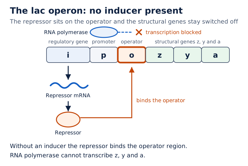
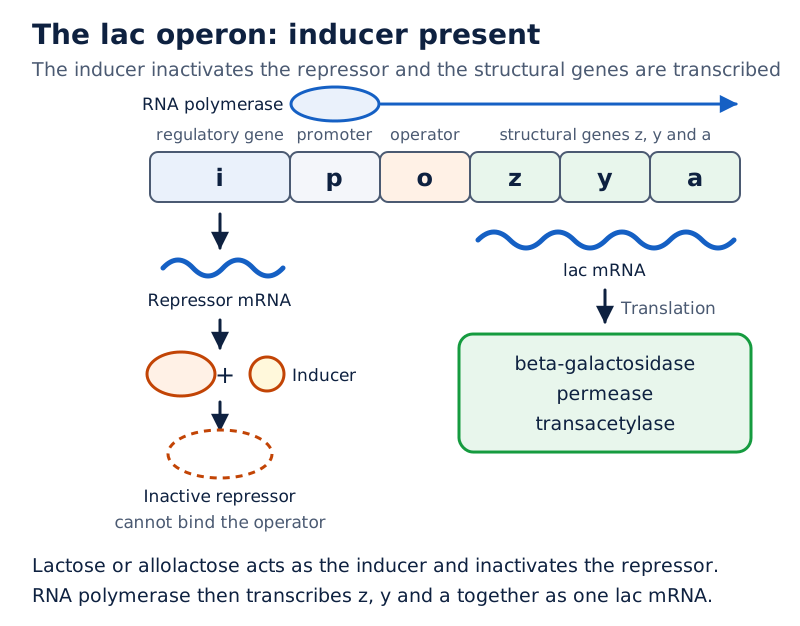
The upper panel shows a repressor bound at the operator when inducer is absent. The lower panel shows inducer interacting with the repressor, followed by lac mRNA formation and production of beta-galactosidase, permease and transacetylase.
Drawn by One Young India.
What did the Human Genome Project investigate?
A genome is an organism’s complete genetic material. The Human Genome Project, abbreviated HGP, began in 1990 and was completed in 2003. Its scale encouraged development of bioinformatics, the use of computational tools to store, retrieve and analyse biological information.
What were its goals and approaches?
The goals included identifying approximately 20,000 to 25,000 genes, determining the sequence of three billion DNA base pairs, storing information in databases, improving analysis tools, transferring related technologies to other sectors, and addressing ethical, legal and social issues.
The Expressed Sequence Tags approach, abbreviated ESTs, focused on identifying genes expressed as RNA. Sequence annotation involved sequencing the whole genome, including coding and non-coding regions, and subsequently assigning functions to different regions.
- Isolate cellular DNA and divide it into smaller random fragments suitable for sequencing, or determining the order of bases.
- Clone the fragments in suitable hosts. Cloning produces many copies; bacteria and yeast were commonly used hosts.
- Sequence the fragments with automated sequencers based on Frederick Sanger's method.
- Use overlapping regions and specialised computer programs to assemble the fragment sequences, then annotate and assign them to chromosomes.
What were the reported findings and applications?
The project-era observations included 3164.7 million bp, an average gene length of 3000 bases, and great variation in gene size. The largest known human gene at the time, dystrophin, spanned 2.4 million bases. Almost all, 99.9 per cent, of human nucleotide bases were the same.
The reported estimate was 30,000 genes, distinct from the project-goal range. Chromosome 1 had most genes (2968), and the Y chromosome had the fewest (231). Functions were unknown for over 50 per cent of discovered genes. Less than 2 per cent of the genome coded for proteins, while repetitive sequences formed a very large portion.
Repeated sequences were thought to have no direct coding functions, but helped explain chromosome structure, dynamics and evolution. About 1.4 million sites of single nucleotide polymorphism, or SNPs, were identified: locations where individuals differed at a single base.
Genome information can help locate disease-associated sequences, trace human history and develop new approaches to diagnosis and treatment. Whole-genome analysis permits study of many genes together. Ethical, legal and social questions concern how genetic information is collected, interpreted and used.
How does DNA fingerprinting distinguish individuals?
DNA fingerprinting compares variable DNA regions to distinguish individuals. Repetitive DNA contains short sequences repeated many times. Some repetitive DNA separates as small peaks from the main DNA peak during density-gradient centrifugation and is called satellite DNA.
These sequences normally do not code for proteins and show a high degree of polymorphism, inherited variation at the genetic level. Their variability makes them useful even though most human DNA sequences are shared.
What is the basis of a DNA fingerprint?
Variable Number of Tandem Repeats, abbreviated VNTR, are mini-satellite DNA sequences whose short repeat units lie one after another. The number of repeats varies, producing DNA fragments of different lengths. Alec Jeffreys initially developed DNA fingerprinting using such sequences.
A probe is labelled DNA used to detect a complementary DNA sequence. Hybridisation means pairing between complementary nucleic-acid sequences. A VNTR probe reveals a characteristic pattern of bands from separated DNA fragments.
- Isolate DNA from the biological sample.
- Digest it with restriction endonucleases, enzymes that cut DNA at specific recognition sequences.
- Separate fragments by electrophoresis, movement through a medium under an electric field.
- Transfer the separated fragments to a synthetic membrane such as nylon or nitrocellulose. This transfer is called blotting.
- Allow a radioactively labelled VNTR probe to hybridise with complementary sequences on the membrane.
- Detect the labelled fragments by autoradiography, recording the pattern produced by their radioactivity.
This describes the earlier Southern-blot hybridisation method. The band pattern differs between individuals except monozygotic twins, identical twins arising from the same fertilised egg. Variation inherited from parents also makes the technique useful in establishing biological parentage.
What are its uses and ethical concerns?
Applications include forensic identification, identification for legal investigations, disputed paternity and studies of population and genetic diversity. DNA from different tissues of one individual shows the same degree of polymorphism, enabling comparisons between biological samples.
Ethical concerns include informed consent, agreement after understanding the proposed use of a sample, and genetic privacy, protection against unauthorised access to genetic information. Results may reveal sensitive family relationships; access and use should therefore be carefully controlled.
Glossary
- Nucleotide — The repeating unit of a nucleic acid, containing a nitrogenous base, pentose sugar and phosphate group.
- Complementarity — Specific pairing of bases that allows one nucleic-acid strand to specify the sequence of another.
- Nucleosome — A repeating chromatin unit formed by DNA wrapped around a positively charged histone octamer.
- Transformation — An inherited change produced when genetic material from one bacterial strain changes the characteristics of another.
- Semiconservative replication — DNA copying in which each daughter molecule retains one parental strand and contains one newly synthesised strand.
- Promoter — A DNA sequence providing the binding site for RNA polymerase at the beginning of a transcription unit.
- Cistron — A segment of DNA that contains the information specifying a single polypeptide chain.
- Intron — An intervening sequence present in a primary transcript but removed during processing to form mature RNA.
- Exon — A sequence retained in mature RNA after the removal of introns and joining of remaining sequences.
- Codon — A sequence of three successive mRNA nucleotides specifying an amino acid or a stop signal.
- Anticodon — A triplet of bases in tRNA that pairs with a complementary codon in messenger RNA.
- Operon — A group of structural genes whose transcription is regulated together through a common promoter and control sequences.
- Inducer — A substance that permits expression of an inducible operon by interacting with and inactivating its repressor.
- Sequence annotation — Assignment of functions to different regions of a genome after its nucleotide sequence has been determined.
- DNA polymorphism — Inherited variation in DNA sequences that occurs at appreciable frequency within a population.
Common errors and misconceptions
- Misconception: Griffith proved that DNA was the transforming substance. Correct: He demonstrated transformation but did not establish the substance's biochemical identity; Avery, MacLeod and McCarty identified DNA.
- Misconception: Heavy nitrogen in the Meselson-Stahl experiment was radioactive. Correct: ¹⁵N was a non-radioactive isotope, and DNA samples were distinguished by density.
- Misconception: Discontinuous DNA synthesis occurs in the 3′ to 5′ direction. Correct: New DNA synthesis proceeds 5′ to 3′ on both templates; ligase joins discontinuously synthesised fragments.
- Misconception: The coding DNA strand is the strand copied during transcription. Correct: RNA polymerase copies the template strand; the RNA resembles the coding strand with U replacing T.
- Misconception: Degeneracy means one codon specifies several amino acids. Correct: It means that some amino acids have more than one codon; the code is nearly universal, not completely universal.
- Misconception: Introns remain in mature mRNA and exons are removed. Correct: Splicing removes introns and joins exons in a defined order.
- Misconception: The lac operon has absolutely no expression without lactose. Correct: A very low level of expression must remain so that permease is available for lactose entry.
- Misconception: DNA fingerprints distinguish every pair of people. Correct: The characteristic pattern differs between individuals except monozygotic twins.
Exam-style questions with model answers
Q1. Distinguish a nucleoside from a nucleotide by composition. [2 marks]
- A nucleoside consists of a nitrogenous base joined to a pentose sugar, without a phosphate group.
- A nucleotide contains a nitrogenous base, pentose sugar and phosphate group; it is formed by adding phosphate to a nucleoside.
Q2. In a transformation experiment, purified DNA from smooth, virulent bacteria transforms living rough bacteria. Protein-digesting and RNA-digesting enzymes do not stop transformation, but DNA-digesting enzyme does. Explain the four findings. [4 marks]
- Transformation by purified DNA shows that material in this DNA preparation can transfer the information needed for the rough bacteria to acquire the smooth form.
- Persistence after protein digestion indicates that protein is not the transforming substance responsible for the observed change.
- Persistence after RNA digestion similarly indicates that RNA is not required as the transforming substance in this experiment.
- Loss of transformation after DNA digestion identifies DNA as the essential transforming material, linking the inherited change specifically to DNA.
Q3. E. coli with both DNA strands labelled with non-radioactive heavy nitrogen, ¹⁵N, is transferred to medium containing light nitrogen, ¹⁴N. Assume semiconservative replication, a 20-minute generation time and no return to heavy medium. Predict the DNA-density classes after 20 and 40 minutes, giving the strand composition and proportions. [4 marks]
- After 20 minutes, one generation has passed. All extracted DNA has intermediate density, producing a hybrid band rather than separate heavy and light bands.
- Each hybrid molecule contains one heavy parental strand and one newly synthesised light strand. The light medium supplies the nitrogen incorporated into the new strands.
- After 40 minutes, two generations have passed. DNA occurs in two density classes, hybrid and light, with equal amounts in each class and no completely heavy DNA.
- Hybrid molecules retain a heavy strand paired with a light strand. Light molecules contain two light strands, formed when first-generation light strands serve as templates.
Q4. The template strand of a transcription unit is 3′-ATGCATGCATGCATGCATGCATGC-5′. During transcription, RNA is synthesised 5′ to 3′; template A pairs with RNA U, T with A, G with C and C with G. Write the RNA sequence, explain its direction, and state its relationship to the coding DNA strand. [3 marks]
- The RNA sequence is 5′-UACGUACGUACGUACGUACGUACG-3′, obtained by replacing each template base with its stated RNA complement.
- The RNA runs antiparallel to the supplied template. Because the template is written 3′ to 5′ from left to right, the new RNA is written 5′ to 3′.
- The coding DNA strand has the same base sequence and direction as the RNA, except that thymine occupies the positions where RNA contains uracil.
Q5. Explain splicing, capping and tailing during the conversion of eukaryotic hnRNA into mature mRNA. State the end modified in each end-modification process. [3 marks]
- Splicing removes the introns from the primary RNA transcript and joins the retained exons in a defined order, producing a continuous processed sequence.
- Capping adds the unusual nucleotide methyl guanosine triphosphate to the 5′ end of the hnRNA during its processing.
- Tailing adds 200 to 300 adenylate residues to the 3′ end without using a DNA template, forming the poly-A tail.
Q6. Describe translation from amino-acid activation to release of the completed polypeptide, including the roles of tRNA, ribosomes and the stop signal. [5 marks]
- Amino acids are activated using ATP and attached to their corresponding tRNAs. This charging, or aminoacylation, prepares amino acids for addition to the growing chain.
- Translation begins when a ribosome binds mRNA at the start codon AUG. An initiator tRNA recognises this codon and establishes initiation.
- During elongation, charged tRNAs successively recognise mRNA codons through complementary pairing of their anticodons, bringing the required amino acids into position.
- The ribosome brings amino acids together, peptide bonds form, and it advances from codon to codon. Bacterial rRNA catalyses peptide-bond formation.
- At a stop codon, a release factor terminates translation and releases the complete polypeptide. No tRNA carrying an additional amino acid corresponds to that stop signal.
Q7. Explain how the lac operon responds when lactose becomes available to E. coli in the absence of a preferred carbon source such as glucose. Include the initial repressed condition and the need for basal expression. [5 marks]
- Initially, the i gene continuously produces a repressor. Without an inducer, this protein binds the operator and prevents transcription of the operon's structural genes.
- A very low basal level of expression remains necessary. It supplies permease, allowing lactose to enter the bacterial cell when lactose becomes available.
- Lactose or allolactose acts as an inducer by interacting with the repressor and inactivating it, removing the repression of transcription.
- RNA polymerase can then transcribe the structural genes z, y and a together, forming the messenger RNA used to produce their proteins.
- The products are beta-galactosidase, permease and transacetylase. Beta-galactosidase splits lactose into glucose and galactose; permease increases entry of beta-galactosides, supporting use of the available lactose.
Q8. Describe the six main steps of the earlier Southern-blot method of DNA fingerprinting using a radioactively labelled VNTR probe. [6 marks]
- Isolate DNA from the biological sample so that its variable repeat-containing regions can be examined by the subsequent fragment-separation procedure.
- Digest the DNA with restriction endonucleases, enzymes that recognise particular DNA sequences and cut the molecules into fragments suitable for comparison.
- Separate the resulting DNA fragments by electrophoresis, producing an arrangement of fragments that can be transferred and probed while preserving their separation.
- Transfer the separated fragments to a synthetic membrane such as nylon or nitrocellulose. This transfer of DNA is the blotting step.
- Hybridise the membrane-bound DNA with the radioactively labelled VNTR probe. The probe pairs with complementary sequences among the separated DNA fragments.
- Detect the hybridised fragments by autoradiography. The labelled fragments produce bands, and their arrangement gives the characteristic DNA-fingerprinting pattern for comparison.
Key takeaways
- DNA has antiparallel, complementary strands; specific base pairing explains both its double-helical structure and its capacity to direct copying.
- Griffith demonstrated transformation, Avery and colleagues identified DNA as the transforming material, and Hershey and Chase traced DNA into bacteria.
- Semiconservative replication gives every daughter DNA molecule one parental strand and one newly synthesised strand.
- Transcription copies a DNA template into RNA; eukaryotic messenger RNA requires splicing, capping and tailing before translation.
- The genetic code is triplet, degenerate and nearly universal; start and stop signals define the translated region.
- Transfer RNA matches codons to amino acids, while ribosomes organise their joining into a polypeptide chain.
- The lac operon links lactose availability to enzyme synthesis through an inducer interacting with a repressor.
- Genome sequencing investigates the complete DNA sequence, whereas DNA fingerprinting compares highly variable regions for identification and relatedness.
Test yourself
What does “antiparallel” mean in a DNA double helix?
The two strands run in opposite directions: one runs 5′ to 3′ and the other 3′ to 5′.
Why do histones associate readily with DNA?
Histones carry positive charges, whereas DNA is negatively charged, allowing DNA to wrap around histone cores.
Which enzyme treatment stopped transformation in Avery and colleagues' investigation?
DNase treatment stopped transformation because it digested the DNA responsible for transferring the inherited characteristic.
Why are fragments produced on one side of a replication fork?
The templates are antiparallel, but DNA polymerase synthesises new DNA only 5′ to 3′, causing discontinuous synthesis on one template.
How do exons differ from introns after RNA processing?
Exons remain joined in mature RNA, whereas introns are removed during the process of splicing.
Why can several codons specify the same amino acid?
There are 61 amino-acid-specifying codons but only 20 amino acids, so some amino acids must be specified by more than one codon. This redundancy is called degeneracy of the genetic code.
What happens to the lac repressor when it interacts with an inducer?
The repressor becomes inactive, permitting RNA polymerase to transcribe the structural genes of the operon.
What do ESTs and sequence annotation investigate differently?
ESTs focus on genes expressed as RNA; sequence annotation assigns functions after sequencing coding and non-coding genome regions.
Which exception limits identification by the described VNTR band patterns?
Monozygotic twins share the same characteristic VNTR band pattern, so this method cannot distinguish them.
Write about it
Turn a question from this chapter into a paper.
The Writing Studio has a structure guide, a rubric and citation tools for 15 formats, from essays to research papers. Drafting is free with an account. Learnacy Essential adds feedback from an AI writing coach and submission to The OYI Review.
Learnacy Essential is ₹1,499 for the year. Publication depends on review and is not guaranteed.