
Breathing and exchange of gases | ISC Class 11 Biology Notes
On this page
This note covers respiratory organs in animals, the human respiratory system, breathing movements, pulmonary volumes and capacities, gaseous exchange, oxygen and carbon dioxide transport, the oxyhaemoglobin dissociation curve, chloride shift, regulation of breathing and respiratory disorders.
What connects breathing with cellular respiration?
Cells require a continuous supply of oxygen (O₂) and removal of carbon dioxide (CO₂). Oxygen is used indirectly in breaking down simple substances such as glucose, amino acids and fatty acids to obtain energy. Carbon dioxide is released during these breakdown reactions.
Catabolism means the breakdown of substances in cells. Cellular respiration involves oxygen utilisation for these reactions and the resulting release of carbon dioxide. Moving air into the lungs is one step in the wider sequence that connects the atmosphere with cells.
Definition: Breathing, or pulmonary ventilation, brings atmospheric air into the lungs and releases carbon dioxide-rich air from them. Pulmonary means relating to the lungs.
What are the steps in the complete sequence?
- Ventilation: atmospheric air enters the lungs, and air rich in carbon dioxide leaves them.
- Alveolar exchange: gases diffuse across the membrane of the alveoli, the small air sacs in the lungs. Diffusion is movement down a concentration or pressure gradient.
- Blood transport: blood carries oxygen towards body tissues and carbon dioxide towards the lungs.
- Tissue exchange: oxygen and carbon dioxide diffuse between blood and tissues, which are groups of cells performing related functions.
- Cellular use: cells use oxygen in catabolic reactions and release carbon dioxide.
The sequence explains why air movement and gas transport must be considered together. Ventilation supplies the exchange surface; diffusion transfers gases across it; blood connects the lungs to the tissues. Cellular reactions provide the continuing need for oxygen delivery and carbon dioxide removal.
Inspiration is the entry of atmospheric air into the lungs. Expiration is the release of alveolar air. These alternating movements renew lung air, while exchange with the blood depends on differences in the pressures of individual gases.
Which respiratory organs occur in different animals?
Animal breathing mechanisms vary mainly with habitat, the environment in which an animal lives, and level of organisation, the complexity of its body structure. Some animals exchange gases over their body surface, while others have specialised structures for this purpose.
How do the main respiratory surfaces compare?
| Animal or group | Respiratory surface or organ | Important feature |
|---|---|---|
| Sponges, coelenterates and flatworms | Entire body surface | Exchange by simple diffusion |
| Earthworms | Moist cuticle | The cuticle is the outer body covering used for exchange |
| Insects | Tracheal tubes | A network of tubes transports atmospheric air within the body |
| Most aquatic arthropods and molluscs | Gills | Specialised structures supplied with blood vessels |
| Fishes | Gills | Gills provide the respiratory organs among these vertebrates |
| Amphibians, reptiles, birds and mammals | Lungs | Lungs provide organs for gaseous exchange |
| Frogs | Moist skin as well as lungs | Skin provides an additional respiratory surface |
Branchial respiration means respiration through gills. Pulmonary respiration means respiration through lungs. Gills are vascularised structures, meaning they contain a blood supply; lungs are vascularised sacs. These terms identify the organ involved in exchange.
Cutaneous respiration means respiration through the skin. Amphibians such as frogs can use their moist skin as well as their lungs. Therefore, identifying lungs in an amphibian does not exclude the skin from its respiratory activity.
Keep the qualification most aquatic arthropods and molluscs when associating these groups with gills. The description is not an unrestricted claim about every aquatic animal. Likewise, insect tracheal tubes and the human trachea belong to different respiratory arrangements, despite the similarity of their names.
How is the human respiratory system organised?
The respiratory system includes air passages and two lungs. Air enters through the external nostrils, passes along the nasal passage into the nasal chamber, and reaches the pharynx. Part of the pharynx is a common passage for food and air.
The pharynx opens through the larynx into the trachea, the main air tube. The larynx is a cartilaginous sound box. Cartilage is the supporting tissue forming this box and the rings that support several air passages.
The glottis, the opening into the larynx, can be covered during swallowing by the epiglottis, a thin, elastic cartilaginous flap. This prevents food from entering the larynx. The flap and the sound box therefore have different functions.
How do the air passages branch?
The trachea divides at the level of the fifth thoracic vertebra, a bone of the chest portion of the backbone, into right and left primary bronchi. A bronchus is one of these branching air passages. Repeated division produces secondary and tertiary bronchi.
Further branches form bronchioles, smaller air passages, ending in thin terminal bronchioles. The trachea, primary, secondary and tertiary bronchi, and initial bronchioles have incomplete cartilaginous supporting rings. This support should not be extended to every structure in the lung.
Each terminal bronchiole gives rise to thin, irregular-walled, vascularised sacs called alveoli; one sac is an alveolus. The branching network of bronchi, bronchioles and alveoli forms the lungs. The alveoli provide the principal exchange surfaces.
How do the conducting and exchange parts differ?
The conducting part extends from the external nostrils to the terminal bronchioles. It carries atmospheric air towards the alveoli, removes foreign particles, moistens the air and brings it to body temperature. These functions prepare the air reaching the exchange surface.
The exchange part consists of alveoli and their ducts. Here oxygen and carbon dioxide actually diffuse between blood and air. Thus, an air passage can be essential to respiration without itself being the principal site of gaseous exchange.
What the figure shows
Human respiratory system
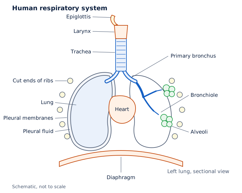
The drawing labels the epiglottis, larynx, trachea, bronchus, lung, bronchiole and alveoli. A sectional view of the left lung shows branching passages. The pleural membranes, pleural fluid, cut ends of ribs, heart and diaphragm are also labelled.
Drawn by One Young India.
How do the chest wall and muscles produce breathing?
The lungs lie in the thoracic chamber, the chest cavity. Its boundaries are the vertebral column behind, the sternum or breastbone in front, the ribs at the sides and the dome-shaped diaphragm, a muscular partition, below.
Each lung has a double covering called the pleura. The outer membrane contacts the thoracic lining; the inner membrane contacts the lung surface. Pleural fluid between them reduces friction on the lung surface.
The thoracic chamber is anatomically airtight. Its arrangement makes changes in chest-cavity volume produce changes in lung volume. Intrapulmonary pressure means pressure within the lungs; atmospheric pressure is the pressure of the surrounding air.
What is the sequence during inspiration?
- The diaphragm contracts, increasing the volume of the thoracic chamber.
- The external intercostal muscles, muscles between the ribs, contract and lift the ribs and sternum.
- Thoracic volume increases, and pulmonary volume increases with it.
- Intrapulmonary pressure falls below atmospheric pressure, creating a pressure gradient, or difference in pressure between the two regions.
- Air moves from outside into the lungs down this pressure gradient.
How does expiration reverse the sequence?
Relaxation of the diaphragm and intercostal muscles returns the diaphragm and sternum to their normal positions. Thoracic and pulmonary volumes decrease. Intrapulmonary pressure rises to slightly above atmospheric pressure, so air is expelled from the lungs.
| Feature | Inspiration | Expiration during ordinary breathing |
|---|---|---|
| Diaphragm | Contracts | Relaxes |
| Ribs and sternum | Are raised by external intercostal contraction | Return to their normal positions |
| Thoracic volume | Increases | Decreases |
| Pulmonary volume | Increases | Decreases |
| Intrapulmonary pressure | Falls below atmospheric pressure | Rises slightly above atmospheric pressure |
| Air movement | Into the lungs | Out of the lungs |
External and internal intercostals are specialised muscles between the ribs involved in generating breathing pressure gradients. Additional abdominal muscles can increase the strength of inspiration and expiration. On average, a healthy human breathes 12 to 16 times per minute.
What the figure shows
Mechanism of breathing
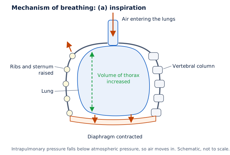
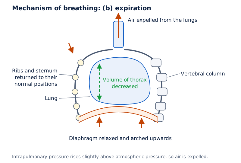
In panel (a), arrows show air entering, raised ribs and sternum, and a contracted diaphragm with increased thoracic volume. Panel (b) shows air leaving, ribs and sternum returning, and a relaxed diaphragm arched upwards with decreased thoracic volume.
Drawn by One Young India.
What do respiratory volumes and lung capacities measure?
Respiratory volumes describe quantities of air associated with particular breathing movements or remaining in the lungs. A spirometer estimates the air volumes involved in breathing movements and helps assess pulmonary function. Volume is expressed here in mL, meaning millilitres.
Which four respiratory volumes must be distinguished?
| Volume and abbreviation | Meaning | Approximate value or average range |
|---|---|---|
| Tidal volume (TV) | Air inspired or expired during a normal breath | Approximately 500 mL |
| Inspiratory reserve volume (IRV) | Additional air inspired by forcible inspiration beyond a normal inspiration | Average 2500 to 3000 mL |
| Expiratory reserve volume (ERV) | Additional air expired by forcible expiration beyond a normal expiration | Average 1000 to 1100 mL |
| Residual volume (RV) | Air remaining even after forcible expiration | Average 1100 to 1200 mL |
The values describe approximate amounts or average ranges, not compulsory values for every individual. At ordinary breathing rates, a healthy man can inspire or expire approximately 6000 to 8000 mL per minute. Tidal volume describes one normal breathing movement.
How are pulmonary capacities constructed?
A pulmonary capacity combines respiratory volumes. The abbreviations in the following relationships name the volumes defined above; the plus sign means addition. Each capacity also has a starting or finishing condition that is essential to its definition.
| Capacity and abbreviation | Definition | Relationship |
|---|---|---|
| Inspiratory capacity (IC) | Total air inspired after a normal expiration | IC = TV + IRV |
| Expiratory capacity (EC) | Total air expired after a normal inspiration | EC = TV + ERV |
| Functional residual capacity (FRC) | Air remaining after a normal expiration | FRC = ERV + RV |
| Vital capacity (VC) | Maximum air inspired after forced expiration, or expired after forced inspiration | VC = ERV + TV + IRV |
| Total lung capacity (TLC) | Total air accommodated at the end of forced inspiration | TLC = RV + ERV + TV + IRV = VC + RV |
Normal expiration leaves functional residual capacity in the lungs, whereas forced expiration still leaves residual volume. Vital capacity measures a maximum movable volume; total lung capacity includes that volume together with the residual air.
Note: Identify whether a definition begins after normal or forced breathing before choosing a capacity. Functional residual capacity includes expiratory reserve volume because that air can still be expelled after a normal expiration.
How do pressure differences drive gas exchange?
Partial pressure is the pressure contributed by one gas in a gas mixture. The symbol pO₂ represents oxygen partial pressure, and pCO₂ represents carbon dioxide partial pressure. Here the letter p denotes partial pressure.
The unit mm Hg means millimetres of mercury, a pressure unit. Oxygenated blood has gained oxygen at the lungs; deoxygenated blood returns after supplying tissues. The word deoxygenated does not mean that the blood contains no oxygen.
What are the partial pressures at the exchange sites?
| Respiratory gas | Atmospheric air, mm Hg | Alveoli, mm Hg | Deoxygenated blood, mm Hg | Oxygenated blood, mm Hg | Tissues, mm Hg |
|---|---|---|---|---|---|
| O₂ | 159 | 104 | 40 | 95 | 40 |
| CO₂ | 0.3 | 40 | 45 | 40 | 45 |
At the lungs, alveolar pO₂ of 104 mm Hg exceeds the 40 mm Hg in arriving deoxygenated blood. Oxygen therefore diffuses into blood. Carbon dioxide diffuses oppositely because its partial pressure is 45 mm Hg in arriving blood and 40 mm Hg in alveoli.
At tissues, oxygenated blood has pO₂ of 95 mm Hg compared with tissue pO₂ of 40 mm Hg, favouring oxygen delivery. Tissue pCO₂ is 45 mm Hg compared with 40 mm Hg in arriving oxygenated blood, favouring carbon dioxide entry into blood.
What else affects diffusion?
Exchange occurs by simple diffusion, mainly according to pressure or concentration gradients. Gas solubility and membrane thickness also affect its rate. Solubility describes how readily a substance dissolves. Carbon dioxide has a solubility 20 to 25 times higher than oxygen.
Consequently, much more carbon dioxide can cross the diffusion membrane per unit difference in partial pressure than oxygen. A smaller carbon dioxide pressure difference should therefore not be interpreted as meaning that carbon dioxide cannot be exchanged effectively.
The diffusion membrane has three major layers: the thin squamous epithelium of the alveolus, the endothelium of the alveolar capillary and the basement substance between them. Squamous epithelium is a lining of flattened cells; endothelium lines blood vessels.
A capillary is a fine blood vessel at an exchange surface. Basement substance includes the thin supporting membranes associated with the alveolar and capillary linings. The total diffusion-membrane thickness is much less than a millimetre.
What the figure shows
Alveolus and pulmonary capillary
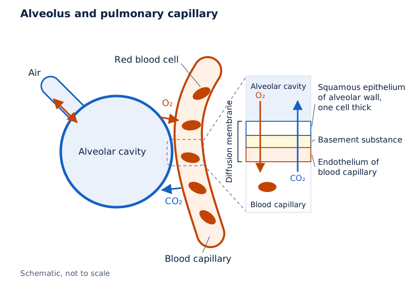
The drawing shows an alveolar cavity beside a blood capillary containing red blood cells. Labels identify one-cell-thick squamous epithelium, basement substance and capillary endothelium. Arrows indicate air movement and oxygen and carbon dioxide exchange.
Drawn by One Young India.
How is oxygen carried and released by blood?
About 97 per cent of oxygen is carried by red blood cells (RBCs), the blood cells containing the respiratory pigment haemoglobin. The remaining 3 per cent is dissolved in plasma, the fluid part of blood.
Haemoglobin is a red, iron-containing pigment. Oxygen binds reversibly with it to form oxyhaemoglobin, the oxygen-bound form. Reversible binding means that oxygen can attach and later be released. Each haemoglobin molecule can carry a maximum of four oxygen molecules.
Which conditions favour loading and unloading?
Oxygen binding depends primarily on pO₂. It is also influenced by pCO₂, temperature and hydrogen ion concentration. H⁺ denotes a hydrogen ion, a positively charged particle; the superscript plus indicates its positive charge.
| Condition | At alveoli | At tissues |
|---|---|---|
| pO₂ | High | Low |
| pCO₂ | Low | High |
| Hydrogen ion concentration | Lesser | High |
| Temperature | Lower | Higher |
| Favoured process | Oxyhaemoglobin formation | Oxygen dissociation from oxyhaemoglobin |
Dissociation means separation of oxygen from its bound form. Conditions at the lung surface favour oxygen loading, whereas tissue conditions favour unloading. Every 100 mL of oxygenated blood can deliver around 5 mL of oxygen under normal physiological conditions.
What does the oxygen dissociation curve show?
The oxygen dissociation curve, also called the oxyhaemoglobin dissociation curve, plots percentage saturation of haemoglobin with oxygen against pO₂. Percentage saturation expresses how fully haemoglobin is loaded with oxygen as a percentage of its oxygen-binding capacity.
The curve is sigmoid, meaning S-shaped. It helps relate oxygen loading to partial pressure and study the influence of factors such as carbon dioxide and hydrogen ions on binding. It is not a plot of breathing rate against time.
What the figure shows
Oxygen dissociation curve
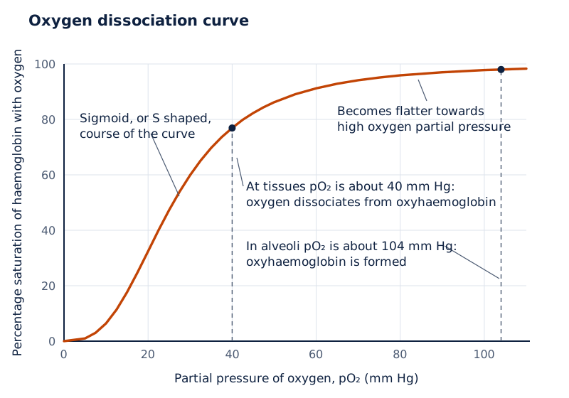
The horizontal axis is oxygen partial pressure in mm Hg; the vertical axis is percentage saturation of haemoglobin with oxygen. The red curve rises in an S-shaped course and becomes flatter towards high oxygen partial pressures.
Drawn by One Young India.
How is carbon dioxide transported, and what is chloride shift?
Blood transports carbon dioxide in three main forms. Bicarbonate, represented by HCO₃⁻, is a negatively charged ion carrying nearly 70 per cent of it. The superscript minus denotes negative charge. About 7 per cent is dissolved in plasma.
About 20 to 25 per cent is carried as carbamino-haemoglobin, carbon dioxide bound to haemoglobin. This must be distinguished from oxyhaemoglobin, which carries oxygen. High pCO₂ and low pO₂ at tissues favour carbon dioxide binding.
Low pCO₂ and high pO₂ at alveoli favour carbon dioxide release from carbamino-haemoglobin. Thus, oxygen and carbon dioxide binding respond to the different conditions at the two ends of the transport route.
How does bicarbonate formation work?
Carbonic anhydrase is an enzyme, a biological catalyst that speeds a reaction. RBCs contain it in very high concentration, while plasma contains minute quantities. It facilitates the reversible conversion associated with carbon dioxide transport.
In the reaction below, H₂O means water and H₂CO₃ means carbonic acid. The double arrow, ⇌, means that the reaction can proceed in either direction. Carbonic acid can separate into bicarbonate and hydrogen ions.
CO₂ + H₂O ⇌ H₂CO₃ ⇌ HCO₃⁻ + H⁺
- Catabolism raises tissue pCO₂, so carbon dioxide diffuses into blood, including RBCs and plasma.
- Carbonic anhydrase facilitates the reaction of carbon dioxide with water, producing carbonic acid.
- Carbonic acid forms bicarbonate and hydrogen ions, allowing carbon dioxide to be carried in bicarbonate form.
- At the alveoli, lower pCO₂ favours the reverse reaction, regenerating carbon dioxide and water.
- The regenerated carbon dioxide is released at the alveoli and can leave with expired air.
Every 100 mL of deoxygenated blood delivers approximately 4 mL of carbon dioxide to the alveoli. This is a delivery amount, distinct from the proportions carried in the three transport forms.
What ion exchange accompanies bicarbonate transport?
Chloride shift is the exchange of bicarbonate and chloride ions across the RBC membrane during carbon dioxide transport. Cl⁻ denotes the chloride ion, a negatively charged ion. This exchange maintains electrical neutrality, or balance of electric charge, in the cell.
At tissues, bicarbonate formed inside RBCs moves out into plasma, while chloride ions move into the cells. This links bicarbonate production in RBCs with its transport in plasma without an unbalanced loss of negative charge from the cells.
At the lungs, the exchange reverses: bicarbonate enters RBCs and chloride leaves. Bicarbonate is then available for conversion back towards carbon dioxide, which diffuses into the alveoli. Remember both the tissue direction and the reverse direction at the lungs.
How is the rhythm of breathing regulated?
The neural system, the body's system of nerve communication, adjusts respiratory rhythm to tissue demands. A respiratory rhythm centre in the medulla, a region of the brain, is primarily responsible for this regulation.
A pneumotaxic centre in the pons, another brain region, can moderate the rhythm centre. Its signals can reduce the duration of inspiration and thereby alter respiratory rate. A change in inspiration duration is therefore part of nervous regulation.
Which chemical changes are detected?
A chemosensitive area, an area responsive to chemical changes, lies beside the rhythm centre. It is highly sensitive to carbon dioxide and hydrogen ions. An increase in these substances can activate it and produce signals to the rhythm centre.
The rhythm centre then makes adjustments in respiration through which these substances can be eliminated. This relationship connects the chemical consequences of tissue activity with changes in breathing. The controlling signal is distinct from the muscle movements that it regulates.
Receptors are structures that detect changes and generate signals. Receptors associated with the aortic arch, the curved portion of the main artery leaving the heart, and the carotid artery, an artery supplying the head, also detect changes in carbon dioxide and hydrogen ion concentration.
These receptors send signals to the respiratory rhythm centre for corrective action. The role of oxygen in regulating respiratory rhythm is quite insignificant. Do not replace this qualified description with the stronger statement that oxygen has absolutely no role.
How can the control system be followed logically?
Separate the detector, coordinating centre and resulting adjustment. The chemosensitive area and associated arterial receptors detect chemical changes. The medullary rhythm centre coordinates regulation, and the pneumotaxic centre can modify its activity by reducing inspiration duration.
How do respiratory disorders affect breathing and exchange?
Respiratory disorders can affect the conducting passages, the alveolar exchange surface or lung tissue exposed to dust. The location and nature of the damage help distinguish asthma, emphysema and occupational respiratory disorders.
What happens in asthma and emphysema?
Asthma involves difficulty in breathing with wheezing, a whistling breathing sound, due to inflammation of the bronchi and bronchioles. Inflammation is a tissue response involving irritation and swelling. The affected structures are part of the branching air passages.
Emphysema is a chronic, or long-lasting, disorder in which alveolar walls are damaged. This decreases the respiratory surface available for gaseous exchange. Cigarette smoking is one of the major causes; that wording does not identify it as the sole cause.
The distinction is structural: asthma concerns inflammation in bronchi and bronchioles, whereas emphysema damages alveolar walls. Both are respiratory disorders, but describing them simply as breathing difficulty would omit the different underlying changes.
Why can occupational dust damage lungs?
In certain industries, especially grinding and stone-breaking, the quantity of dust can exceed what the body's defence mechanisms can fully handle. Occupational means associated with a person's work. The exposure is therefore related to conditions in the workplace.
Long exposure can cause inflammation leading to fibrosis, the proliferation of fibrous tissue, and serious lung damage. Fibrosis describes a tissue change rather than the dust itself. Workers in such industries should wear protective masks.
Preserve the conditional sequence: long dust exposure can produce inflammation and fibrosis. The explanation does not mean that every brief contact with dust necessarily produces permanent lung damage. The important link is prolonged occupational exposure and the resulting risk to lung tissue.
Glossary
- Pulmonary ventilation — Movement of atmospheric air into the lungs and carbon dioxide-rich alveolar air out.
- Alveolus — A thin-walled, vascularised air sac forming a principal site of pulmonary gas exchange.
- Pleura — Double membrane covering the lungs, with friction-reducing pleural fluid between its layers.
- Inspiration — Entry of atmospheric air when pressure within the lungs falls below atmospheric pressure.
- Tidal volume — Volume of air inspired or expired during a normal breathing movement.
- Residual volume — Air remaining in the lungs even after a forcible expiration has occurred.
- Vital capacity — Maximum air breathed out after forced inspiration, or breathed in after forced expiration.
- Partial pressure — Pressure contributed by an individual gas within a mixture of gases.
- Oxyhaemoglobin — The reversible combination formed when oxygen binds to haemoglobin in red blood cells.
- Carbamino-haemoglobin — Haemoglobin combined with carbon dioxide, accounting for one form of carbon dioxide transport.
- Carbonic anhydrase — Enzyme present abundantly in red blood cells that facilitates reversible conversion of carbon dioxide and water into carbonic acid.
- Chloride shift — Exchange of bicarbonate and chloride ions across red blood cell membranes during carbon dioxide transport.
- Pneumotaxic centre — Centre in the pons whose signals can shorten inspiration and modify respiratory rate.
- Fibrosis — Proliferation of fibrous tissue that can follow inflammation caused by prolonged occupational dust exposure.
Common errors and misconceptions
- Misconception: Air enters because pressure inside the lungs rises. Correct: Inspiration follows a fall in intrapulmonary pressure below atmospheric pressure.
- Misconception: Every air passage is an exchange surface. Correct: The conducting part carries and conditions air; alveoli and their ducts form the exchange part.
- Misconception: Forced expiration empties the lungs completely. Correct: Residual volume remains even after forcible expiration.
- Misconception: Vital capacity and total lung capacity are identical. Correct: Total lung capacity includes residual volume in addition to vital capacity.
- Misconception: Most carbon dioxide travels as carbamino-haemoglobin. Correct: Nearly 70 per cent travels as bicarbonate, while about 20 to 25 per cent travels as carbamino-haemoglobin.
- Misconception: Chloride leaves RBCs when bicarbonate leaves them at tissues. Correct: Chloride enters as bicarbonate leaves; the exchange reverses at the lungs.
- Misconception: Asthma and emphysema involve identical structural damage. Correct: Asthma involves inflamed bronchi and bronchioles; emphysema damages alveolar walls and reduces respiratory surface.
Exam-style questions with model answers
Q1. Distinguish the conducting part from the exchange part of the human respiratory system by their extent and function. [2 marks]
- The conducting part extends from the nostrils to terminal bronchioles; it transports, cleans, moistens and warms incoming air.
- The exchange part consists of alveoli and their ducts, where oxygen and carbon dioxide diffuse between air and blood.
Q2. Explain normal inspiration through five successive changes, beginning with muscle action and ending with airflow. [5 marks]
- The diaphragm contracts, increasing thoracic chamber volume and beginning the mechanical changes that allow the lungs to expand during inspiration.
- The external intercostal muscles contract and raise the ribs and sternum, contributing to the increase in the volume of the chest cavity.
- The anatomical arrangement of the lungs within the thorax causes pulmonary volume to increase along with the increased thoracic volume.
- This increase in pulmonary volume lowers intrapulmonary pressure below atmospheric pressure, establishing a pressure difference between the surrounding air and lungs.
- Air consequently moves from the atmosphere into the lungs down this pressure gradient, producing the inward airflow called inspiration.
Q3. Tidal volume is 500 mL, inspiratory reserve volume is 2500 mL, expiratory reserve volume is 1000 mL and residual volume is 1100 mL. Calculate inspiratory capacity, functional residual capacity and vital capacity, stating each relationship. [3 marks]
- Inspiratory capacity equals tidal volume plus inspiratory reserve volume: 500 + 2500 = 3000 mL, the air inspired after normal expiration.
- Functional residual capacity equals expiratory reserve volume plus residual volume: 1000 + 1100 = 2100 mL, the air remaining after normal expiration.
- Vital capacity equals expiratory reserve volume plus tidal volume plus inspiratory reserve volume: 1000 + 500 + 2500 = 4000 mL.
Q4. At the lungs, oxygen partial pressure is 104 mm Hg in alveoli and 40 mm Hg in arriving blood; carbon dioxide partial pressure is 40 mm Hg in alveoli and 45 mm Hg in arriving blood. Predict each gas's diffusion direction and explain both using the supplied values. [2 marks]
- Oxygen diffuses from alveoli into blood, down its partial-pressure gradient from 104 mm Hg to 40 mm Hg.
- Carbon dioxide diffuses from blood into alveoli, down its partial-pressure gradient from 45 mm Hg to 40 mm Hg.
Q5. Define the oxyhaemoglobin dissociation curve, state its shape, and contrast the conditions favouring oxygen loading at alveoli with unloading at tissues. [4 marks]
- The curve plots percentage saturation of haemoglobin with oxygen against oxygen partial pressure, showing the relationship between oxygen availability and haemoglobin loading.
- It has a sigmoid, or S-shaped, course, rising and then becoming flatter towards higher oxygen partial pressures.
- At alveoli, high oxygen partial pressure, low carbon dioxide partial pressure, lesser hydrogen ion concentration and lower temperature favour oxyhaemoglobin formation.
- At tissues, low oxygen partial pressure, high carbon dioxide partial pressure, high hydrogen ion concentration and higher temperature favour oxygen dissociation.
Q6. Explain carbon dioxide transport as bicarbonate, including carbonic anhydrase, chloride shift at tissues, and reversal at the lungs. [5 marks]
- Carbon dioxide produced by tissue catabolism diffuses into blood because tissue carbon dioxide partial pressure is high; it enters both red blood cells and plasma.
- Carbonic anhydrase, abundant in red blood cells, facilitates carbon dioxide combination with water; the resulting carbonic acid forms bicarbonate and hydrogen ions.
- At tissues, bicarbonate leaves red blood cells for plasma while chloride enters them. This chloride shift maintains electrical neutrality as negative ions are exchanged.
- At the lungs, bicarbonate moves back into red blood cells while chloride leaves. The reversal supplies bicarbonate for conversion back towards carbon dioxide.
- Low alveolar carbon dioxide partial pressure favours the reverse chemical reaction, forming carbon dioxide and water. Carbon dioxide diffuses into alveoli for expiration.
Q7. Explain the roles of the respiratory rhythm centre, pneumotaxic centre, chemosensitive area and arterial receptors in respiratory regulation. [4 marks]
- The respiratory rhythm centre in the medulla is primarily responsible for regulating the breathing rhythm according to the demands of body tissues.
- The pneumotaxic centre in the pons moderates rhythm-centre activity; its signals can shorten inspiration and thereby alter the respiratory rate.
- The adjacent chemosensitive area responds strongly to carbon dioxide and hydrogen ions, signalling the rhythm centre when their concentrations increase.
- Receptors associated with the aortic arch and carotid artery detect carbon dioxide and hydrogen ion changes and send signals for corrective respiratory adjustments.
Q8. Distinguish asthma, emphysema and occupational respiratory disorders by their characteristic tissue changes; include the stated major cause or exposure where applicable. [3 marks]
- Asthma involves inflammation of the bronchi and bronchioles, producing difficulty in breathing with wheezing; the affected structures are conducting air passages.
- Emphysema is a chronic disorder in which damaged alveolar walls reduce respiratory surface. Cigarette smoking is one of its major causes.
- Prolonged exposure to heavy occupational dust, especially in grinding or stone-breaking, can cause inflammation followed by fibrosis and serious lung damage.
Key takeaways
- Respiration links ventilation, alveolar diffusion, blood transport, tissue diffusion and cellular oxygen utilisation in a continuous functional sequence.
- Inspiration increases thoracic and pulmonary volumes, lowering intrapulmonary pressure below atmospheric pressure so air enters.
- Normal expiration leaves functional residual capacity, while forcible expiration still leaves residual volume inside the lungs.
- Partial-pressure gradients favour oxygen movement from alveoli to tissues and carbon dioxide movement in the opposite direction.
- Haemoglobin loads oxygen under alveolar conditions and releases it under tissue conditions; its oxygen dissociation curve is sigmoid.
- Most carbon dioxide travels as bicarbonate; chloride shift exchanges bicarbonate and chloride across red blood cell membranes.
- The medullary rhythm centre regulates breathing, with modification by the pneumotaxic centre and chemical detection systems.
- Asthma affects bronchi and bronchioles, emphysema damages alveolar walls, and prolonged occupational dust exposure can cause fibrosis.
Test yourself
Which additional respiratory surface can frogs use besides lungs?
Frogs can exchange gases through their moist skin; this is called cutaneous respiration.
What does pleural fluid do?
It reduces friction on the lung surface between the two pleural membranes.
Why does increased pulmonary volume cause inspiration?
It lowers intrapulmonary pressure below atmospheric pressure, so air moves into the lungs.
Which two volumes make up functional residual capacity?
Expiratory reserve volume and residual volume together form functional residual capacity.
How many oxygen molecules can one haemoglobin molecule carry?
Each haemoglobin molecule can carry a maximum of four oxygen molecules.
What happens to chloride during bicarbonate export from RBCs at tissues?
Chloride enters the red blood cells as bicarbonate moves into plasma, maintaining electrical neutrality.
How can the pneumotaxic centre alter respiratory rate?
Its signals can reduce the duration of inspiration and thereby alter the respiratory rate.
Which structural change reduces the exchange surface in emphysema?
Damage to alveolar walls decreases the respiratory surface available for gaseous exchange.
Write about it
Turn a question from this chapter into a paper.
The Writing Studio has a structure guide, a rubric and citation tools for 15 formats, from essays to research papers. Drafting is free with an account. Learnacy Essential adds feedback from an AI writing coach and submission to The OYI Review.
Learnacy Essential is ₹1,499 for the year. Publication depends on review and is not guaranteed.