
Nobel Prize in Chemistry 2008: The Green Fluorescent Protein GFP
On this page
What was the Nobel Prize in Chemistry 2008 awarded for?
The Royal Swedish Academy of Sciences gave the Nobel Prize in Chemistry 2008 jointly to three scientists "for the discovery and development of the green fluorescent protein, GFP".
It names the exact achievement being honoured.
In plain language, the prize rewards two linked things. First, it rewards finding a protein inside a jellyfish that glows bright green under ultraviolet or blue light without needing any extra chemical fuel.
Second, it rewards turning that protein into a working tool that other scientists could attach to any protein of interest, so that they could watch it glow and therefore see exactly where it goes and what it does inside a living cell.
The prize's full official name is the Nobel Prize in Chemistry, awarded by the Royal Swedish Academy of Sciences.
It was announced on 8 October 2008, and the prize amount that year was 10,000,000 Swedish kronor, shared equally among the three laureates.
The Academy described GFP as a "guiding star for biochemistry" in its press release, because the glowing protein let researchers watch processes inside cells that used to be completely invisible.
Who are the laureates?
All three laureates received an equal one third share of the prize, reflecting the fact that the discovery and development of GFP happened in three connected stages, each carried out by a different scientist.
Osamu Shimomura
Osamu Shimomura was born on 27 August 1928 in Kyoto, Japan, and died on 19 October 2018 in Nagasaki, Japan.
At the time of the award he was affiliated with the Marine Biological Laboratory (MBL) in Woods Hole, Massachusetts, USA, and with Boston University Medical School. He held one third of the prize.
Shimomura was the scientist who actually found and purified GFP from the jellyfish Aequorea victoria, and he later worked out the chemical structure of the part of the protein responsible for its glow.
Martin Chalfie
Martin Chalfie was born on 15 January 1947 in Chicago, Illinois, USA. At the time of the award he was affiliated with Columbia University in New York. He held one third of the prize.
Chalfie's key contribution was showing that GFP could glow inside other living organisms without any help from jellyfish machinery, and he used it as a genetic tag to light up specific cells in the roundworm Caenorhabditis elegans.
Roger Y. Tsien
Roger Y. Tsien was born on 1 February 1952 in New York, USA, and died on 24 August 2016 in Eugene, Oregon, USA.
At the time of the award he was affiliated with the University of California, San Diego, and with the Howard Hughes Medical Institute. He held one third of the prize.
Tsien explained the chemistry of how GFP's glow forms, and then re-engineered the protein to create a whole range of new colours beyond green.
What problem were scientists trying to solve?
Through the twentieth century, chemists and biologists learned a great deal about the molecules that make up a living cell: the structure of proteins, the sequence of DNA, and the chemical pathways that keep cells alive.
But none of this knowledge gave them a simple way to watch a particular protein moving and working inside a living, intact cell in real time.
Tens of thousands of different proteins exist inside any organism, each doing a specific job, and when these proteins malfunction, illness often follows.
Scientists wanted to know, for example, which cells in a growing embryo would become insulin-producing beta cells in the pancreas, how nerve cells develop and wire themselves together in the brain, or how cancer cells spread through the body.
Ordinary microscopes could not reveal these processes because individual proteins inside a transparent, colourless cell are simply invisible.
What was needed was a built-in light, something that could be attached to any protein the researcher cared about and that would glow on its own, visible under a microscope, without disturbing the normal behaviour of the cell.
This is exactly the gap that the green fluorescent protein filled, and the story of the 2008 prize is the story of how that gap was closed, from a chance observation in a Japanese laboratory to a tool now used in laboratories across the world.
How does GFP actually glow?
GFP comes from the jellyfish Aequorea victoria, which lives off the west coast of North America and glows green along the edge of its umbrella-shaped body when disturbed. The protein itself is a chain of 238 amino acids.
Three of these amino acids, in positions 65 to 67, fold up inside the protein in a way that lets them react spontaneously with each other to form a small light-absorbing and light-emitting unit called a chromophore.
What makes this chromophore remarkable is that it needs no other protein or enzyme to form: it only needs ordinary molecular oxygen.
This is very different from many other glowing biological molecules, which depend on a continuous supply of extra chemical fuel.
Once the chromophore has formed, shining ultraviolet or blue light onto the protein excites the chromophore, and it then releases that extra energy as green light.
- The GFP gene is switched on inside a cell, and the cell's protein-making machinery builds the 238-amino-acid chain.
- The chain folds into its natural barrel-shaped structure, bringing three particular amino acids close together.
- These three amino acids react with each other and, in the presence of oxygen, form the chromophore.
- Shining blue or ultraviolet light on the folded protein excites the chromophore.
- The chromophore releases the absorbed energy as green fluorescent light, which a microscope can detect.
Diagram
the GFP barrel and its chromophore
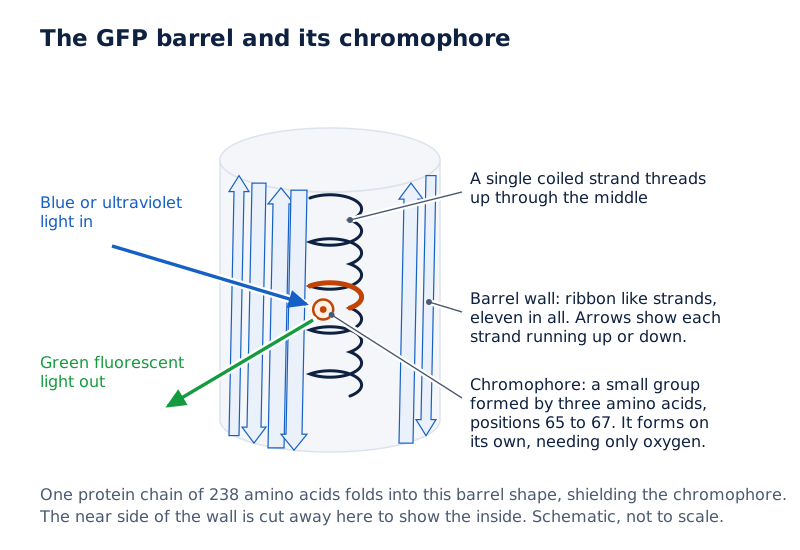
Draw a short, closed cylinder made of ribbon-like strands running up and down its sides, representing the protein's outer shell.
Inside the cylinder, draw a single coiled strand running through the middle, and mark a small highlighted group on this inner strand as the chromophore, protected inside the barrel.
Drawn by One Young India.
Because the chromophore sits inside a protective barrel-shaped shell of the protein, it is shielded from the surrounding cell and keeps glowing reliably, which is one reason GFP turned out to be such a dependable tool.
How did Chalfie turn GFP into a genetic tag?
Finding a glowing protein in a jellyfish is one thing; making it useful to thousands of other researchers is another.
The breakthrough came when scientists realised that the gene for GFP could be copied from the jellyfish and inserted into the DNA of a completely different organism, where it would still make a working, glowing protein.
Martin Chalfie worked with the small roundworm Caenorhabditis elegans, a transparent worm with only 959 cells that is widely studied because scientists can see its internal organs directly through an ordinary microscope.
Chalfie obtained the cloned GFP gene and attached it to a genetic "switch", called a promoter, that is normally active only in six specific touch-sensing nerve cells of the worm.
When this combined piece of DNA was placed inside the worm, those six nerve cells, and only those cells, began to glow bright green under ultraviolet light, because only in those cells was the switch turned on.
This showed that GFP could be used as a genetic tag: wherever a researcher placed the GFP gene behind a chosen switch or attached it to a chosen protein, that location would light up and could be followed under a microscope.
Chalfie's results, published in the journal Science in February 1994, demonstrated that GFP glowed without needing any other protein from the jellyfish, which meant it would work in essentially any living organism.
Why this mattered for biology
Once GFP could be fused to the gene of any protein a scientist wanted to study, researchers could watch that protein's exact location, its movement around the cell and its interactions with other proteins, all while the cell stayed alive and otherwise unharmed.
How did Tsien expand GFP into a rainbow of colours?
Roger Tsien's contribution began with explaining the underlying chemistry of GFP's glow, including confirming that the chromophore forms by itself once oxygen is available, without needing an unknown jellyfish enzyme as many scientists had assumed.
This explained why GFP worked so reliably in bacteria, worms and other organisms far removed from its natural jellyfish home.
Tsien then used genetic engineering to swap individual amino acids within the GFP protein, which changed the wavelengths of light the protein could absorb and emit.
Through many such changes, his laboratory produced versions of GFP that glowed more brightly, lasted longer under the microscope, and shone in new colours such as cyan, blue and yellow.
One colour Tsien could not get directly from GFP was red, which is especially useful because red light travels further through living tissue than green light.
Researchers Mikhail Matz and Sergei Lukyanov found a naturally red fluorescent protein, called DsRed, in a coral. DsRed worked poorly as a tag because it only functioned as a bulky cluster of four protein chains.
Tsien's group redesigned DsRed so that it worked as a single, well-behaved chain, and from this redesigned protein they created a family of new red and orange fluorescent proteins.
| Protein or colour | Origin | Contribution |
|---|---|---|
| Green fluorescent protein (GFP) | Jellyfish Aequorea victoria | Original glowing protein isolated by Shimomura |
| Cyan, blue and yellow GFP variants | Engineered from GFP | Developed by Tsien's laboratory by changing amino acids |
| DsRed | Coral Discosoma | Natural red protein found by Matz and Lukyanov |
| mPlum, mCherry, mStrawberry, mOrange, mCitrine | Redesigned from DsRed | Single-chain coloured proteins created by Tsien's group |
With proteins now available across the whole visible spectrum, researchers could tag several different proteins or cell types at once, each in its own colour, and follow them simultaneously under the microscope.
How did the discovery unfold?
The path from a glowing jellyfish to a universal laboratory tool took nearly five decades and passed through the hands of all three laureates in turn.
| Year | Event |
|---|---|
| 1955 | Osamu Shimomura joins Professor Hirata's laboratory at Nagoya University and studies the glowing ostracod Cypridina. |
| 1960 | Shimomura joins Frank Johnson's laboratory at Princeton University to study the bioluminescent jellyfish Aequorea victoria. |
| 1961 | Shimomura and Johnson collect jellyfish at Friday Harbor, Washington, and isolate a blue-glowing protein later named aequorin. |
| 1962 | Shimomura and Johnson publish the first description of a second, greenish, fluorescent protein from the jellyfish, later called GFP. |
| 1979 | Shimomura identifies the chemical structure of the light-emitting chromophore inside GFP. |
| 1988 | Martin Chalfie first hears about GFP at a seminar at Columbia University and sees its potential as a biological marker. |
| 1992 | Douglas Prasher clones the full gene for GFP, and Chalfie expresses the gene successfully in the bacterium E. coli. |
| 1994 | Chalfie publishes results showing GFP glowing in the touch receptor neurons of the roundworm C. elegans; Tsien explains that the chromophore forms using only oxygen. |
| 1999 | Mikhail Matz and Sergei Lukyanov discover the red fluorescent protein DsRed in a coral; Tsien's group later redesigns it for use as a tag. |
| 2008 | The Nobel Prize in Chemistry is awarded jointly to Shimomura, Chalfie and Tsien for the discovery and development of GFP. |
Why does it matter?
GFP and its coloured relatives became what the Nobel Committee called a guiding star for biochemistry because they let researchers watch processes that were previously hidden inside living cells.
By attaching the GFP gene to the gene of a protein under study, scientists can follow that protein's movements, its position inside the cell and its interactions with other molecules, all without killing or seriously disturbing the cell.
The press release noted that GFP has been used to track nerve cell damage in Alzheimer's disease, to watch how insulin-producing beta cells form in a developing pancreas, and to observe how cancer cells spread.
In one striking experiment described on the Nobel website, researchers coloured different nerve cells in a mouse's brain with varying amounts of yellow, cyan and red fluorescent proteins, producing a multicoloured map of the brain's nerve network that they called the "brainbow".
Beyond pure research, fluorescent proteins have practical biotechnology uses too. Scientists have engineered bacteria that glow green in the presence of arsenic, a serious problem in drinking water in parts of South-East Asia, and other organisms that fluoresce when exposed to the explosive TNT or to heavy metals such as cadmium and zinc.
One open question the Nobel material raises is why Aequorea victoria evolved GFP and aequorin at all, since no one has established what advantage the jellyfish itself gains from glowing.
How does this connect to what you study?
GFP sits at the meeting point of several ideas taught in school biology and chemistry: how genes code for proteins, how proteins fold into specific three-dimensional shapes, and how light interacts with molecules through absorption and emission.
The idea that a gene can be copied from one organism and inserted into another, called gene cloning, is central to the GFP story and to modern genetic engineering more broadly.
The GFP chromophore's dependence on oxygen also illustrates a basic chemistry idea, that a reaction needing a simple small molecule like oxygen can produce a complex, useful outcome once the right structural conditions (the folded protein shape) are in place.
Students studying cell biology encounter the same model organisms used in the GFP story, including the bacterium Escherichia coli and the roundworm Caenorhabditis elegans, both widely used because they are easy to grow and study in a laboratory.
Quick facts for exams
The Nobel Prize in Chemistry 2008 was awarded jointly to Osamu Shimomura, Martin Chalfie and Roger Y. Tsien "for the discovery and development of the green fluorescent protein, GFP".
It was announced by the Royal Swedish Academy of Sciences on 8 October 2008. Shimomura, working in Japan and later at Princeton, first isolated GFP from the jellyfish Aequorea victoria.
Chalfie showed GFP could glow as a genetic tag in bacteria and in the roundworm C. elegans. Tsien explained the chemistry of GFP's glow and engineered a rainbow of new fluorescent colours, including red variants derived from a coral protein.
Each laureate received an equal one third share of the prize, worth 10,000,000 Swedish kronor in total.
| Fact | Detail |
|---|---|
| Prize | Nobel Prize in Chemistry 2008 |
| Date announced | 8 October 2008 |
| Laureates | Osamu Shimomura, Martin Chalfie, Roger Y. Tsien |
| Countries of birth | Japan (Shimomura), USA (Chalfie and Tsien) |
| Affiliations at award | MBL and Boston University Medical School (Shimomura); Columbia University (Chalfie); University of California, San Diego, and Howard Hughes Medical Institute (Tsien) |
| Shares | One third each |
| Citation | "for the discovery and development of the green fluorescent protein, GFP" |
| Prize amount | 10,000,000 Swedish kronor |
Note: Source. The prize facts in this note are from the Nobel Prize's official site, nobelprize.org.
Glossary
- Green fluorescent protein (GFP) — a protein from the jellyfish Aequorea victoria that glows green under ultraviolet or blue light.
- Chromophore — the small chemical group inside a protein that absorbs and then releases light energy, producing fluorescence.
- Fluorescence — the process by which a molecule absorbs light of one colour and emits light of a different, usually longer, wavelength.
- Bioluminescence — the production of light by a living organism through a chemical reaction, as seen in aequorin.
- Aequorin — a blue-light-emitting protein from Aequorea victoria, discovered before GFP by Shimomura and Johnson.
- Genetic tag — a marker, such as GFP, attached to a gene or protein so its location and activity can be tracked.
- Promoter — a region of DNA near a gene that switches the gene on or off in particular cells.
- Gene cloning — copying a gene from one organism's DNA and inserting it into another organism so it can be studied or used.
- Amino acid — one of the small building-block molecules that link together in chains to form proteins.
- Caenorhabditis elegans — a small, transparent roundworm widely used as a model organism in biology.
- Escherichia coli (E. coli) — a common intestinal bacterium often used as a simple test organism in genetic engineering.
- DsRed — a naturally red fluorescent protein found in coral, later redesigned by Tsien's laboratory for use as a tag.
- Model organism — a simple, well-studied species used by researchers to understand processes common to many living things.
Common errors and misconceptions
- Misconception: GFP needs an extra chemical injected into the cell to glow. Correct: GFP's chromophore forms on its own once oxygen is present, and the protein then glows simply when exposed to blue or ultraviolet light.
- Misconception: Shimomura discovered GFP's use as a laboratory tool. Correct: Shimomura isolated and characterised GFP itself; it was Chalfie who first showed it could work as a genetic tag in other organisms.
- Misconception: GFP only ever glows green. Correct: Engineered variants developed mainly by Tsien and others glow in colours across the visible spectrum, including blue, cyan, yellow, orange and red.
- Misconception: The red fluorescent protein DsRed came directly from GFP. Correct: DsRed is a naturally occurring red protein found in a coral by Matz and Lukyanov, later redesigned by Tsien's group.
- Misconception: All three laureates worked together as a single team. Correct: They worked independently across different decades and institutions, each building on the previous laureate's findings.
- Misconception: GFP is only useful in jellyfish. Correct: GFP has been expressed successfully in bacteria, yeast, worms, fruit flies and mammalian cells, which is exactly why it became a universal tool.
- Misconception: The prize was given for curing a specific disease. Correct: The prize was given for a research tool; the citation itself only credits the discovery and development of GFP, not any single medical application.
Exam-style questions with model answers
Q1. In which year was the Nobel Prize in Chemistry 2008 announced? [1 mark]
- It was announced on 8 October 2008 by the Royal Swedish Academy of Sciences.
Q2. Name the three laureates of the Nobel Prize in Chemistry 2008. [2 marks]
- The laureates were Osamu Shimomura, Martin Chalfie and Roger Y. Tsien, who shared the prize equally.
Q3. Explain why GFP's chromophore is described as needing no auxiliary factor to form. [4 marks]
- Three amino acids near positions 65 to 67 in the folded GFP protein react spontaneously with each other to form the light-emitting chromophore.
- This reaction requires only ordinary molecular oxygen, which is present in almost all living cells, rather than any special enzyme unique to the jellyfish.
- Because of this, GFP can fold and glow correctly in organisms completely unrelated to jellyfish, such as bacteria, worms and mammalian cells.
- This property is exactly what made GFP useful as a universal genetic tag across the biological sciences.
Q4. Describe Martin Chalfie's key experiment with Caenorhabditis elegans and explain its significance. [4 marks]
- Chalfie attached the cloned GFP gene to a genetic switch that is normally active only in six touch receptor neurons of the roundworm C. elegans.
- When this construct was placed inside the worm, exactly those six nerve cells glowed bright green under ultraviolet light.
- This showed that GFP did not need any other protein from its native jellyfish to glow, and that it could be targeted to specific cells using a chosen gene switch.
- The result, published in Science in 1994, proved that GFP could serve as a universal genetic marker in living organisms far removed from Aequorea victoria.
Q5. Discuss the contributions of each of the three 2008 Chemistry laureates and explain how their work built on each other. [6 marks]
- Osamu Shimomura isolated GFP from the jellyfish Aequorea victoria during research begun in the 1960s, discovering that it glowed bright green under ultraviolet light, and he later worked out the chemical structure of its light-emitting chromophore.
- Martin Chalfie, after learning about GFP at a 1988 seminar, obtained a cloned copy of the GFP gene and demonstrated in the early 1990s that the protein could glow when expressed in the bacterium E. coli and, more strikingly, in specific nerve cells of the roundworm C. elegans, proving GFP could act as a genetic tag in any organism.
- Roger Y. Tsien explained the underlying chemistry showing that the chromophore forms using only oxygen, and he then re-engineered GFP to create brighter, more stable variants glowing in new colours, as well as helping redesign a naturally red coral protein, DsRed, into a usable single-chain tag.
- Together, their work turned a jellyfish curiosity into a universal toolkit that lets researchers watch proteins and cells at work inside living organisms, which is why the Nobel Committee honoured all three jointly for the discovery and development of GFP.
Q6. State the official citation for the Nobel Prize in Chemistry 2008. [1 mark]
- The citation reads "for the discovery and development of the green fluorescent protein, GFP".
Q7. Give one example of a disease process that researchers have studied using GFP. [2 marks]
- Researchers have used GFP to track nerve cell damage during Alzheimer's disease by tagging and following affected neurons under the microscope.
Q8. Why was finding a red fluorescent protein particularly valuable to researchers? [3 marks]
- Red light penetrates biological tissue more easily than green or blue light.
- This makes red fluorescent proteins especially useful for studying cells and organs deep inside a living body rather than only cells near the surface.
- The natural red protein DsRed, found in coral, was redesigned by Tsien's group into a practical single-chain tag to meet this need.
Key takeaways
- The 2008 Chemistry prize went jointly to Shimomura, Chalfie and Tsien for discovering and developing GFP.
- GFP was first isolated from the jellyfish Aequorea victoria by Osamu Shimomura.
- GFP's chromophore forms spontaneously, needing only oxygen and no extra enzyme.
- Martin Chalfie proved GFP could work as a genetic tag in bacteria and in the roundworm C. elegans.
- Roger Y. Tsien explained GFP's chemistry and engineered new colours, including red variants from coral protein DsRed.
- GFP lets scientists watch proteins, cells and processes that were previously invisible inside living organisms.
- Applications include studying Alzheimer's disease, cancer spread, and detecting arsenic or heavy metals in water.
- One open question remains: why Aequorea victoria itself evolved the ability to glow.
Test yourself
Who first isolated GFP, and from which organism?
Osamu Shimomura first isolated GFP from the jellyfish Aequorea victoria during research that began in the 1960s.
What organism did Martin Chalfie use to show GFP could act as a genetic tag?
Martin Chalfie used the roundworm Caenorhabditis elegans, lighting up six specific touch receptor neurons with GFP.
What single small molecule does GFP's chromophore need in order to form?
GFP's chromophore needs only ordinary molecular oxygen to form; no other enzyme or chemical is required.
Which laureate developed a palette of new colours from GFP?
Roger Y. Tsien developed a palette of new fluorescent colours by changing amino acids within the GFP protein.
Where was the coral-derived red protein DsRed originally found?
DsRed was found in the coral Discosoma by researchers Mikhail Matz and Sergei Lukyanov.
When was the 2008 Nobel Prize in Chemistry announced?
The prize was announced by the Royal Swedish Academy of Sciences on 8 October 2008.
Name one real-world use of GFP mentioned in the Nobel material besides basic research.
Scientists have engineered bacteria that glow green in the presence of arsenic, helping detect it in drinking water.