
Anatomy of Flowering Plants | CBSE Class 11 Biology Notes
On this page
Watch & explore
Start with a few high-quality watches, then dive into the notes below.
This note covers the internal organisation of flowering plants, the three tissue systems, the outer covering and its specialised cells, conducting bundles, root and stem structure, leaf anatomy, and the structural differences between the two groups of flowering plants.
What does plant anatomy reveal about tissue organisation?
Plant anatomy is the study of the internal structure of plants. Cells are the basic units of a plant. They form tissues, organised groups of cells, which in turn form organs, structural units such as roots, stems and leaves. Roots, stems and leaves differ in the arrangement of their internal tissues.
Angiosperms are flowering plants. Dicotyledonous plants or dicots have two cotyledons, while monocotyledonous plants or monocots have one. Cotyledons are seed leaves. These two groups show anatomical differences. Comparing the same organ in these groups reveals differences in the type, number and position of conducting bundles.
How are tissue systems classified?
A tissue system groups tissues according to their structure and location in the plant body. The location of a tissue is also related to its function. The three systems are the epidermal, ground and vascular tissue systems.
| Tissue system | Meaning and components | Position or role |
|---|---|---|
| Epidermal tissue system | The outer covering, including epidermal cells and their specialised structures | Forms the outermost covering of the whole plant body |
| Ground or fundamental tissue system | All tissues apart from the epidermis and vascular bundles | Forms the main bulk of the plant |
| Vascular or conducting tissue system | The conducting tissues called xylem and phloem | Conducts water, minerals and food material |
Xylem and phloem are the two conducting tissues that together form a vascular bundle. Their arrangement differs between roots, stems and leaves. A bundle is therefore a structural unit within the vascular tissue system, rather than a separate fourth tissue system.
Plant tissues carry out food formation and storage, transport of water, minerals and food material, and mechanical support. Internal structures also show adaptations to different environments. Anatomy connects the position and structure of tissues with these functions.
Note: A tissue system and an organ are different levels of organisation. A root, stem or leaf contains tissues belonging to different tissue systems; its identity does not come from a single tissue alone.
How does the epidermal tissue system cover the plant?
The epidermis is the outermost layer of the primary plant body, the body formed during initial growth. Its elongated, closely arranged cells form a continuous covering. It is usually single-layered. Its cells are parenchymatous, belonging to parenchyma, a simple tissue. A simple tissue consists of similar cells; thin-walled parenchymatous cells also form inner layers of roots described below.
Epidermal cells have a small amount of cytoplasm, the semi-fluid material within a cell, lining the cell wall. A vacuole is a membrane-bound space in the cytoplasm; in these cells it is large. These features describe the cells forming the covering.
What is the cuticle?
The cuticle is a thick, waxy layer often present outside the epidermis. It prevents water loss. The cuticle is absent in roots. The epidermal covering therefore differs according to the organ being considered.
The epidermal tissue system includes epidermal cells, stomata, which are structures with pores in the leaf epidermis, and epidermal appendages, meaning projections such as hairs. The structures around stomatal pores regulate their opening and closing.
How do root hairs and trichomes differ?
Root hairs are unicellular, meaning single-celled, extensions of epidermal cells. They help absorb water and minerals from the soil. Their connection with absorption differs from the role of the hairs on the shoot.
Trichomes are epidermal hairs on the stem. In the shoot system, the above-ground portion of the plant, they are usually multicellular, meaning made of several cells. They may be branched or unbranched, soft or stiff, and may even be secretory, releasing substances.
Trichomes help prevent water loss through transpiration, the loss of water as vapour from the aerial parts of the plant. Thus, an epidermal projection on a root and a hair on a stem need not have the same cellular organisation or function.
| Feature | Root hairs | Trichomes |
|---|---|---|
| Location | Root epidermis | Stem epidermis and shoot system |
| Cellular organisation | Unicellular epidermal extensions | Usually multicellular in the shoot system |
| Described role | Absorption of water and minerals | Helping prevent water loss through transpiration |
How are stomata and the stomatal apparatus organised?
A single stoma consists of two guard cells enclosing a stomatal pore, also called the stomatal aperture. The pore is the opening between these cells. Stomata regulate both transpiration and gaseous exchange, the exchange of gases.
Guard cells are the paired cells that control stomatal opening and closing. They are bean-shaped; in grasses, they are dumb-bell shaped. Their shape must therefore be described with reference to the plant group rather than treated as identical in all plants.
How do guard-cell walls differ?
The outer walls, facing away from the stomatal pore, are thin. The inner walls, facing towards it, are highly thickened. Guard cells possess chloroplasts, the cell structures associated with photosynthesis, or food formation using light.
Sometimes, a few epidermal cells near the guard cells become specialised in shape and size. These are subsidiary cells. The stomatal aperture, guard cells and surrounding subsidiary cells together form the stomatal apparatus.
Definition: The stomatal apparatus comprises the stomatal aperture, its guard cells and the surrounding subsidiary cells. The pore alone is not the complete apparatus.
What should a labelled stomatal drawing show?
What the figure shows
Stomatal apparatus
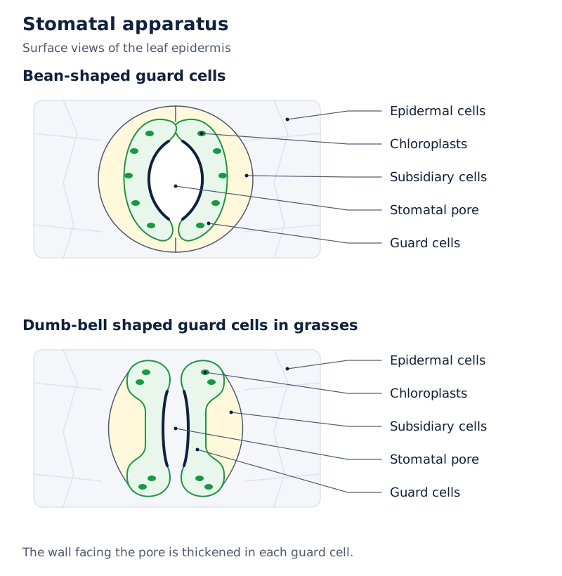
Two drawings compare bean-shaped guard cells with dumb-bell shaped guard cells. Labels identify epidermal cells, subsidiary cells, chloroplasts, guard cells and the stomatal pore. In both drawings, the pore lies between the paired guard cells.
Drawn by One Young India. See Fig. 6.1 in your NCERT textbook
In a labelled drawing, distinguish the surrounding epidermal cells from the guard cells immediately enclosing the pore. Place the chloroplast label within a guard cell. The wall towards the pore and the wall away from it must also remain distinct in the description.
The roles of the different components should remain separate. The guard cells regulate opening and closing. The stomata regulate transpiration and gaseous exchange. Subsidiary cells are identified by their specialised shape and size near the guard cells.
What forms the ground and vascular tissue systems?
The ground tissue comprises all tissues other than the epidermis and vascular bundles. It contains simple tissues such as parenchyma, collenchyma, the supporting tissue found below the epidermis of young dicot stems, and sclerenchyma, the tissue found beneath the outer covering and around conducting bundles in monocot stems.
Parenchymatous cells are usually present in the cortex, the region inside the outer covering, the pericycle, a tissue region internal to the cortex, the central pith, and medullary rays, radial bands of cells between vascular bundles, in primary roots and stems.
In leaves, the ground tissue is called mesophyll. It consists of thin-walled cells containing chloroplasts. Mesophyll occupies the region between the upper and lower epidermis and carries out photosynthesis.
What makes a vascular bundle open or closed?
Cambium is the tissue between xylem and phloem that can produce secondary xylem and phloem. Secondary tissues are the additional tissues formed through this activity. Dicot stems have cambium within their vascular bundles.
An open vascular bundle contains cambium and can form secondary xylem and phloem. A closed vascular bundle lacks cambium and does not form secondary tissues. The vascular bundles of monocotyledons are closed.
How do radial and conjoint arrangements differ?
A radius is a line extending from the centre towards the outside of a section. In a radial arrangement, xylem and phloem alternate along different radii, as in roots. In a conjoint arrangement, they occur together along the same radius.
Conjoint bundles are common in stems and leaves. They usually have phloem located only on the outer side of the xylem. The terms radial and conjoint concern relative position; open and closed concern the presence or absence of cambium.
| Bundle term | Defining feature | Location or consequence |
|---|---|---|
| Radial | Xylem and phloem alternate along different radii | Occurs in roots |
| Conjoint | Xylem and phloem lie together along the same radius | Common in stems and leaves |
| Open | Cambium lies between xylem and phloem | Can form secondary tissues, as in dicot stems |
| Closed | Cambium is absent from the bundle | Does not form secondary tissues, as in monocot bundles |
What the figure shows
Types of vascular bundles
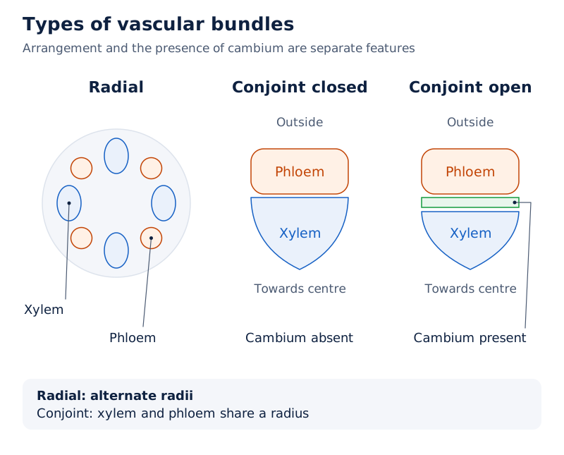
The radial drawing shows alternating xylem and phloem patches. The conjoint closed drawing labels phloem and xylem. The conjoint open drawing adds a cambium band between the phloem and xylem.
Drawn by One Young India. See Fig. 6.2 in your NCERT textbook
How are tissues arranged in a dicot root?
A transverse section, abbreviated as T.S., is a cross-section through an organ. Sections through mature regions help reveal tissue organisation. The sunflower root provides an example of the internal arrangement in a dicot root.
What lies outside the conducting tissues?
The epiblema is the outermost layer of the root. Many of its cells extend outwards as unicellular root hairs. Inside it, the cortex consists of several layers of thin-walled parenchyma with intercellular spaces, or gaps between neighbouring cells.
The endodermis is the innermost cortical layer. In the dicot root, it is a single layer of barrel-shaped cells without intercellular spaces. Both the radial and tangential walls bear deposits of suberin, a waxy material impermeable to water.
Radial walls run in the direction from the centre towards the outside; tangential walls lie across that direction. The suberin deposits form Casparian strips. These strips belong to the endodermal cell walls and must not be confused with the external cuticle.
What lies inside the endodermis?
A few layers of thick-walled parenchymatous cells form the pericycle next to the endodermis. Lateral roots, the branches arising from a root, and vascular cambium begin in these cells during secondary growth, the formation of additional secondary tissues.
The pith is small or inconspicuous. Conjunctive tissue consists of the parenchymatous cells between xylem and phloem. There are usually two to four xylem and phloem patches. Later, a cambium ring develops between these tissues.
The stele includes all tissues on the inner side of the endodermis, including the pericycle, vascular bundles and pith. The endodermis marks its outer boundary; the cortex outside this boundary is not part of the stele.
How can the section be traced from outside to inside?
- Start with the epiblema and its root hairs, which identify the outer surface.
- Move through the several cortical layers and note the spaces between their parenchymatous cells.
- Locate the endodermis at the inner edge of the cortex, followed by the pericycle.
- Examine the xylem and phloem patches, the conjunctive tissue between them, and the small or inconspicuous pith.
This sequence is a route for reading the section, not a sequence in which the tissues develop. It keeps the outer covering, cortical layers and central tissues in their correct spatial relationship.
How does a monocot root compare with a dicot root?
The monocot root resembles the dicot root in many respects. Both contain an outer epidermis, cortex, endodermis, pericycle, vascular bundles and pith. Their differences become clearer when the number of xylem bundles, the pith and secondary growth are compared.
Monocot roots usually have more than six xylem bundles. This condition is called polyarch. Their pith is large and well developed. Monocot roots do not undergo secondary growth.
Which similarities and differences help identify the roots?
| Feature | Dicot root | Monocot root |
|---|---|---|
| Basic tissue regions | Outer covering, cortex, endodermis, pericycle, vascular bundles and pith | The same basic regions are present |
| Conducting tissue arrangement | Xylem and phloem are radial | Xylem and phloem are radial |
| Xylem number | Usually two to four xylem and phloem patches | Usually more than six xylem bundles, described as polyarch |
| Pith | Small or inconspicuous | Large and well developed |
| Secondary growth | Occurs in most dicot roots | Does not occur |
Protoxylem and metaxylem are the earlier-formed and later-formed parts of primary xylem, respectively. In the root drawings, protoxylem is labelled towards the outside of the xylem region, while metaxylem is labelled further inwards.
What the figure shows
Dicot and monocot roots
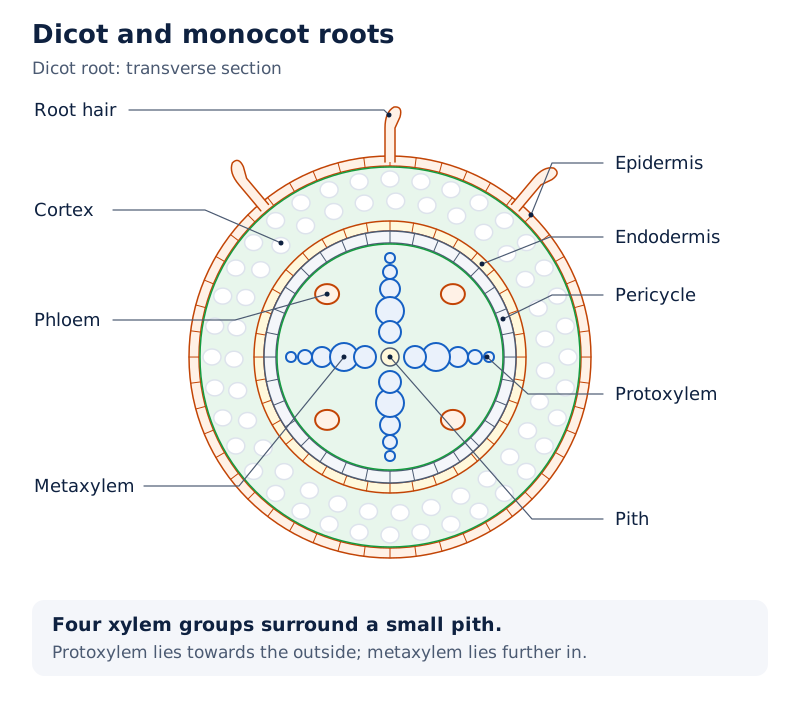
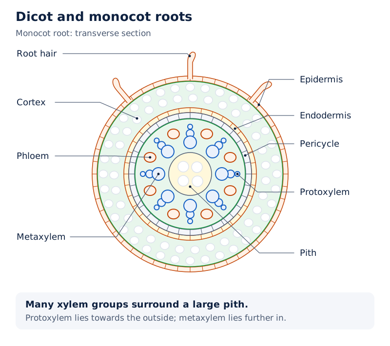
Both sections label root hair, epidermis, cortex, endodermis, pericycle, phloem, protoxylem, metaxylem and pith. The dicot drawing shows fewer xylem groups and a small central pith; the monocot drawing shows more xylem groups surrounding a larger pith.
Drawn by One Young India. See Fig. 6.3 in your NCERT textbook
Root hairs and a cortex occur in both drawings, so their presence alone does not separate the two root types. The useful comparison combines xylem number with pith development. The shared radial organisation identifies a similarity rather than a distinguishing feature.
How are dicot and monocot stems structurally different?
What is the arrangement in a young dicot stem?
The epidermis is the outer protective layer of a typical young dicot stem. A thin cuticle covers it, and it may bear trichomes and a few stomata. The cortex occupies the region between the epidermis and pericycle.
The cortex has three sub-zones. Its outer hypodermis, the region immediately below the epidermis, contains a few layers of collenchymatous cells that give mechanical strength to the young stem. Beneath it are rounded, thin-walled parenchymatous cells with conspicuous intercellular spaces.
The innermost cortical layer is the endodermis. Its cells contain abundant starch grains, so this layer is also called the starch sheath. The pericycle lies inside it, above the phloem, as half-moon-shaped patches of sclerenchyma.
Between the vascular bundles lie a few layers of radially arranged parenchymatous cells forming medullary rays. Numerous vascular bundles form a ring, a characteristic arrangement of the dicot stem. Each bundle is conjoint and open.
The protoxylem is endarch, meaning positioned towards the centre relative to the metaxylem. The central pith contains many rounded parenchymatous cells with large intercellular spaces. The ring of bundles and the central pith must be distinguished in a section.
What changes in a monocot stem?
A monocot stem has a sclerenchymatous hypodermis and many scattered vascular bundles in a large, conspicuous parenchymatous ground tissue. Each bundle has a sclerenchymatous bundle sheath, a layer of tissue surrounding the vascular bundle.
The bundles are conjoint and closed. Peripheral bundles, those near the outside, are generally smaller than central ones. Phloem parenchyma, the parenchymatous component of phloem, is absent. Water-containing cavities occur within the vascular bundles.
| Feature | Dicot stem | Monocot stem |
|---|---|---|
| Hypodermis | Collenchymatous | Sclerenchymatous |
| Bundle distribution | Numerous bundles arranged in a ring | Numerous scattered bundles |
| Relative xylem and phloem position | Conjoint bundles | Conjoint bundles |
| Cambium in bundles | Present, making the bundles open | Absent, making the bundles closed |
| Ground tissue organisation | Distinct cortex, medullary rays and central pith | Large, conspicuous parenchymatous ground tissue |
How can a stem section be examined for identification?
- Locate the vascular bundles and determine whether they form a ring or are scattered through the ground tissue.
- Examine the tissue below the epidermis to distinguish the collenchymatous hypodermis of a dicot stem from the sclerenchymatous hypodermis of a monocot stem.
- Check for cambium between xylem and phloem, identifying open bundles in the dicot stem and closed bundles in the monocot stem.
- Relate the bundle pattern to the surrounding tissues: medullary rays and central pith in the dicot stem, or conspicuous ground tissue and sheathed bundles in the monocot stem.
What the figure shows
Dicot and monocot stems
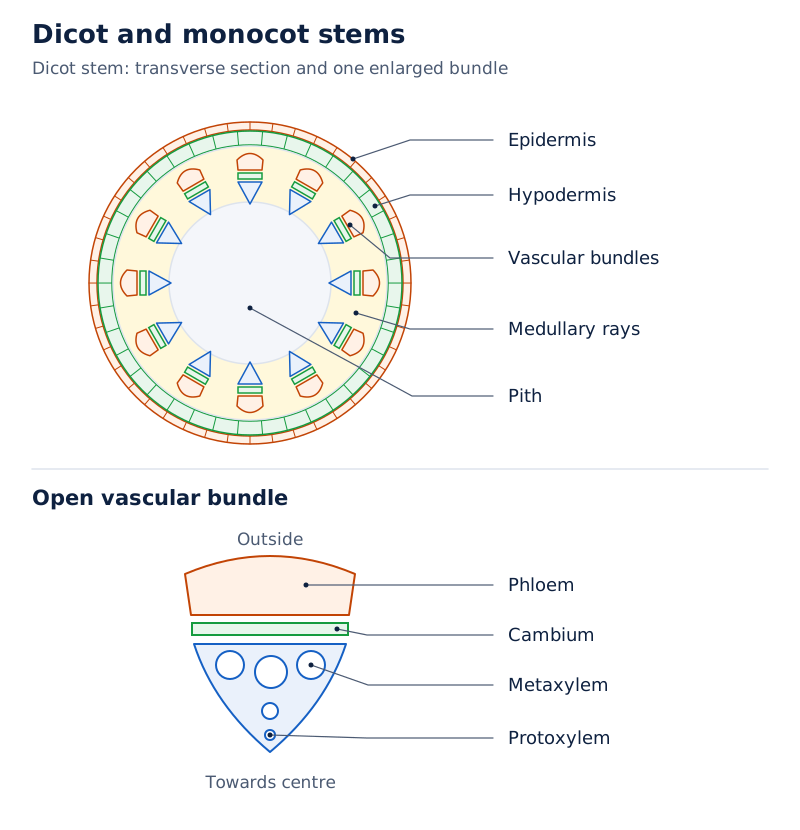
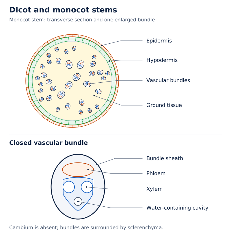
The dicot drawings show a ring of vascular bundles, medullary rays and pith, with an enlarged region labelling phloem, cambium, metaxylem and protoxylem. The monocot drawings show scattered vascular bundles in ground tissue beneath the epidermis and hypodermis.
Drawn by One Young India. See Fig. 6.4 in your NCERT textbook
How is a dorsiventral dicot leaf organised?
A dorsiventral leaf is the dicot leaf type with a differentiated upper and lower internal arrangement. A vertical section through the lamina, or leaf blade, shows three main parts: epidermis, mesophyll and the vascular system.
How do the upper and lower surfaces compare?
The adaxial epidermis covers the upper surface, while the abaxial epidermis covers the lower surface. Both have a conspicuous cuticle. The abaxial epidermis generally bears more stomata than the adaxial epidermis; the adaxial epidermis may even lack stomata.
The mesophyll between these surfaces consists of parenchyma containing chloroplasts and carries out photosynthesis. It is differentiated into two cell types, palisade parenchyma and spongy parenchyma. Their positions, shapes and arrangements differ.
How do the two mesophyll regions differ?
Palisade parenchyma lies on the adaxial side. Its elongated cells stand vertically and parallel to one another. Spongy parenchyma lies below the palisade cells and extends towards the lower epidermis.
Spongy cells are oval or round and loosely arranged. Numerous large spaces and air cavities occur between them. A leaf section therefore shows an upper region of parallel elongated cells and a lower region of loosely arranged cells with conspicuous spaces.
Where are the vascular bundles?
The vascular system occurs in the veins, the conducting strands of the leaf, and the midrib, its main central vein. Bundle size depends on vein size. Dicot leaves have reticulate venation, a network of veins whose thickness varies.
A layer of thick-walled bundle sheath cells surrounds each vascular bundle. In the dicot leaf drawing, xylem lies towards the upper epidermis and phloem towards the lower epidermis. The bundle sheath encloses these conducting tissues within the surrounding mesophyll.
What the figure shows
Dorsiventral leaf
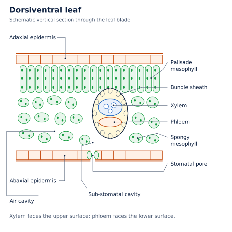
The drawing labels the upper adaxial and lower abaxial epidermis, palisade and spongy mesophyll, air cavity, stomatal pore and the cavity beneath it. A bundle sheath surrounds xylem above phloem in the vascular bundle.
Drawn by One Young India. See Fig. 6.5a in your NCERT textbook
How does an isobilateral leaf differ, and what do bulliform cells do?
An isobilateral leaf is the monocot leaf type with stomata on both epidermal surfaces and mesophyll not differentiated into palisade and spongy parenchyma. Its anatomy resembles a dorsiventral leaf in several ways, but these features distinguish their internal organisation.
What do the veins and mesophyll show?
Monocot leaves have parallel venation, in which veins run parallel to one another. In vertical sections, the vascular bundles have near similar sizes, except in the main veins. This reflects the pattern of venation.
| Feature | Dorsiventral dicot leaf | Isobilateral monocot leaf |
|---|---|---|
| Stomatal distribution | Generally more on the lower epidermis; the upper epidermis may lack them | Present on both epidermal surfaces |
| Mesophyll organisation | Differentiated into palisade and spongy parenchyma | Not differentiated into palisade and spongy parenchyma |
| Venation | Reticulate, with veins varying in thickness | Parallel |
| Vascular bundle size | Depends on the size of the vein | Near similar sizes, except in the main veins |
What the figure shows
Isobilateral leaf
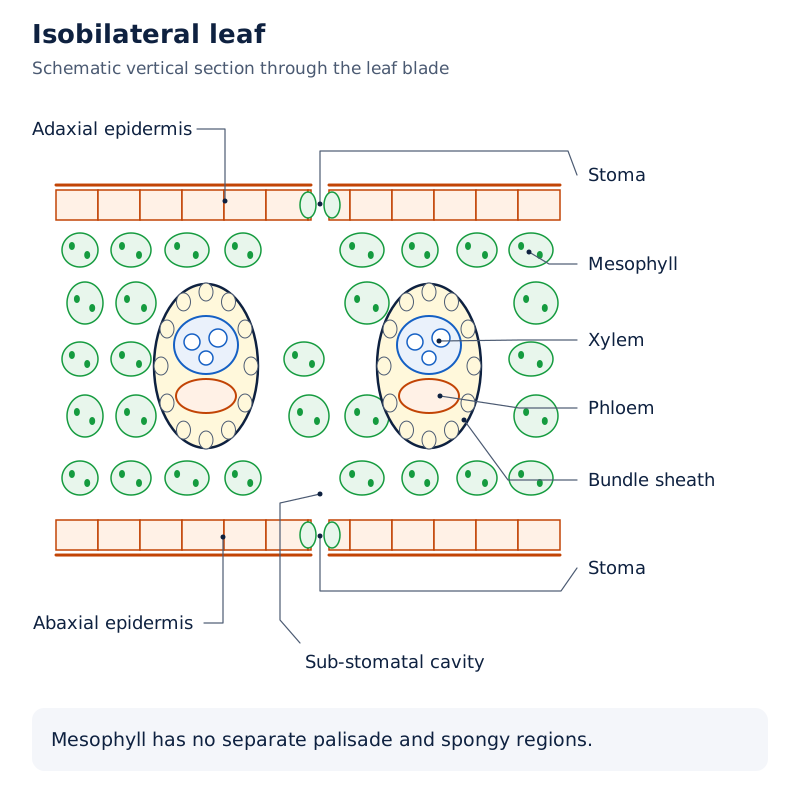
The drawing labels adaxial epidermis, abaxial epidermis, mesophyll, xylem, phloem, stoma and a cavity beneath a stoma. The mesophyll is shown without separate palisade and spongy regions.
Drawn by One Young India. See Fig. 6.5b in your NCERT textbook
How are bulliform cells related to water stress?
In grasses, certain adaxial epidermal cells along the veins become large, empty and colourless. These specialised cells are bulliform cells. Their water condition is associated with whether the leaf surface remains exposed or curls inwards.
- When bulliform cells absorb water, they become turgid, meaning swollen with absorbed water.
- With the bulliform cells turgid, the leaf surface is exposed.
- Under water stress, a shortage of water, the cells become flaccid, meaning they lose their firm, water-filled condition.
- The flaccid cells make the leaf curl inwards, minimising water loss.
Bulliform cells are modified epidermal cells, so they belong to the epidermal tissue system. The mesophyll remains the internal photosynthetic tissue. This distinction connects the position of a cell type with its role without confusing it with the conducting bundles along the veins.
Note: Preserve both qualifications in the comparison: monocot leaf bundles have near similar sizes, with an exception for main veins, and the described bulliform cells occur in grasses.
Glossary
- Anatomy — The study of internal structure, including the arrangement of tissues within plant organs.
- Epidermis — The outermost layer of the primary plant body, usually consisting of a single cell layer.
- Cuticle — A thick, waxy layer often covering the epidermis and preventing loss of water.
- Stomatal apparatus — The stomatal aperture together with its guard cells and surrounding subsidiary cells.
- Trichomes — Epidermal hairs on stems, usually multicellular in the shoot system, that help prevent transpiration-related water loss.
- Ground tissue — All tissues other than the epidermis and vascular bundles, forming the main bulk of the plant.
- Vascular bundle — A structural association of xylem and phloem within the conducting tissue system.
- Open vascular bundle — A bundle containing cambium between xylem and phloem, allowing formation of secondary tissues.
- Endodermis — The innermost layer of the cortex, forming a distinct boundary around the internal tissues.
- Casparian strips — Deposits of water-impermeable suberin on radial and tangential walls of root endodermal cells.
- Stele — All tissues inside the endodermis, including the pericycle, vascular bundles and pith.
- Conjunctive tissue — Parenchymatous cells occupying the regions between xylem and phloem in a dicot root.
- Mesophyll — Chloroplast-containing ground tissue between the upper and lower leaf epidermis that carries out photosynthesis.
- Bulliform cells — Large, empty, colourless cells formed from certain adaxial epidermal cells along the veins in grasses.
Common errors and misconceptions
- Misconception: The epidermis is invariably single-layered and covered by a cuticle. Correct: It is usually single-layered and often covered by a cuticle; the cuticle is absent in roots.
- Misconception: The stomatal pore alone is the stomatal apparatus. Correct: The apparatus includes the aperture, guard cells and surrounding subsidiary cells.
- Misconception: All epidermal hairs are unicellular. Correct: Root hairs are unicellular, while trichomes in the shoot system are usually multicellular.
- Misconception: Open and conjoint mean the same thing. Correct: Open refers to cambium presence; conjoint refers to xylem and phloem sharing the same radius.
- Misconception: A large pith identifies a dicot root. Correct: The dicot root has a small or inconspicuous pith; the monocot root has a large, well-developed pith.
- Misconception: Both stem types have vascular bundles arranged in a ring. Correct: Dicot stems have a ring of bundles, while monocot stems have scattered bundles.
- Misconception: A dorsiventral leaf has stomata exclusively on its lower surface. Correct: The lower epidermis generally has more stomata; the upper epidermis may even lack them.
- Misconception: Turgid bulliform cells curl the leaf inwards. Correct: The surface is exposed when these cells are turgid; flaccidity due to water stress causes inward curling.
Exam-style questions with model answers
Q1. Define the stomatal apparatus and state the role of guard cells. [2 marks]
- The stomatal apparatus consists of the stomatal aperture, the guard cells and the surrounding subsidiary cells.
- Guard cells regulate the opening and closing of stomata, which regulate transpiration and gaseous exchange.
Q2. A stem section has scattered conjoint vascular bundles, each surrounded by a sclerenchymatous sheath, and lacks phloem parenchyma. Identify the stem type and explain your identification using the three observations. [4 marks]
- The section is a monocotyledonous stem, identified from the combination of the supplied features.
- Its scattered conjoint vascular bundles match the bundle distribution of a monocot stem.
- A sclerenchymatous sheath around each vascular bundle is another matching monocot stem feature.
- The stated absence of phloem parenchyma also agrees with monocot stem anatomy, supporting the identification alongside the bundle arrangement and sheath.
Q3. Distinguish radial from conjoint vascular bundles, and open from closed vascular bundles. Give one location for each arrangement or type. [4 marks]
- In radial bundles, xylem and phloem alternate along different radii. This arrangement occurs in roots.
- In conjoint bundles, xylem and phloem lie together along the same radius. Such bundles are common in stems and leaves.
- Open bundles contain cambium between xylem and phloem and can form secondary tissues. They occur in dicot stems.
- Closed bundles lack cambium and do not form secondary tissues. They occur in monocot stems.
Q4. Describe the internal structure of a dicot root under six headings: epiblema, cortex, endodermis, pericycle, conducting region and pith. [6 marks]
- The epiblema forms the outermost layer. Many of its cells extend outwards as unicellular root hairs.
- The cortex consists of several layers of thin-walled parenchymatous cells, with intercellular spaces between neighbouring cells.
- The endodermis is one layer of barrel-shaped cells without intercellular spaces. Suberin forms Casparian strips on radial and tangential walls.
- The pericycle comprises a few layers of thick-walled parenchymatous cells. Lateral roots and vascular cambium originate in these cells during secondary growth.
- The conducting region usually has two to four xylem and phloem patches, with parenchymatous conjunctive tissue between them. A cambium ring develops later.
- The central pith is small or inconspicuous. Along with the pericycle and vascular bundles, it belongs to the stele inside the endodermis.
Q5. Compare dicot and monocot roots in xylem number, pith development and secondary growth. [3 marks]
- A dicot root usually has two to four xylem and phloem patches; a monocot root usually has more than six xylem bundles, the polyarch condition.
- The dicot root pith is small or inconspicuous, whereas the monocot root pith is large and well developed.
- Secondary growth occurs in most dicot roots. Monocot roots do not undergo secondary growth.
Q6. Describe a dorsiventral leaf in five points, covering its epidermis, palisade tissue, spongy tissue, vascular bundle sizes and bundle sheath. [5 marks]
- The adaxial and abaxial epidermis cover the upper and lower surfaces and have a conspicuous cuticle. The lower epidermis generally has more stomata; the upper may lack them.
- The chloroplast-containing mesophyll includes adaxially placed palisade parenchyma. Its elongated cells are arranged vertically and parallel to each other.
- Spongy parenchyma lies below the palisade tissue and extends to the lower epidermis. Its oval or round, loosely arranged cells have numerous large spaces and air cavities between them.
- Vascular bundles occur in the veins and midrib. Their sizes depend on vein size; veins vary in thickness in reticulate venation.
- Each vascular bundle is surrounded by a layer of thick-walled bundle sheath cells, separating the conducting bundle from the surrounding mesophyll.
Q7. In a grass leaf, large, empty, colourless cells occur in the upper epidermis along the veins. Name these cells, describe their water-absorbed state, and explain their response to water stress and its function. [4 marks]
- These modified adaxial epidermal cells are bulliform cells, recognised by the location and appearance given in the question.
- After absorbing water, bulliform cells become turgid, and the leaf surface remains exposed.
- Under water stress, the cells become flaccid and make the leaf curl inwards.
- The inward curling minimises water loss, linking the response of these specialised epidermal cells to the water condition of the leaf.
Q8. Name the three tissue systems of flowering plants and state the components included in each. [3 marks]
- The epidermal tissue system comprises epidermal cells, stomata and epidermal appendages, including root hairs and trichomes.
- The ground tissue system includes all tissues except the epidermis and vascular bundles. It contains simple tissues such as parenchyma, collenchyma and sclerenchyma.
- The vascular tissue system consists of xylem and phloem, the two conducting tissues which together form vascular bundles.
Key takeaways
- The three tissue systems are epidermal, ground and vascular; their structure, position and functions organise the internal plant body.
- Root hairs are unicellular extensions for absorption; shoot trichomes are usually multicellular and help prevent water loss through transpiration.
- The stomatal apparatus includes the aperture, guard cells and surrounding subsidiary cells; guard cells regulate stomatal opening and closing.
- Radial and conjoint describe xylem-phloem arrangement, while open and closed distinguish vascular bundles by cambium presence or absence.
- Dicot roots usually have fewer xylem patches and a small pith; monocot roots usually have more than six xylem bundles and a large pith.
- Dicot stems have ring-arranged open bundles, whereas monocot stems have scattered closed bundles surrounded by sclerenchymatous sheaths.
- Dorsiventral leaves have palisade and spongy mesophyll; isobilateral leaves lack this differentiation and bear stomata on both epidermal surfaces.
- Grass bulliform cells expose the leaf surface when turgid and cause inward curling when flaccid under water stress, minimising water loss.
Test yourself
What makes a bundle open rather than closed?
An open bundle has cambium between xylem and phloem and can form secondary tissues; a closed bundle lacks cambium.
Where are Casparian strips located, and what forms them?
They occur on the radial and tangential walls of root endodermal cells and consist of water-impermeable, waxy suberin.
Which tissues belong to the stele?
All tissues inside the endodermis belong to the stele, including the pericycle, vascular bundles and pith.
Why is the dicot stem endodermis called the starch sheath?
Its cells are rich in starch grains, giving this innermost cortical layer the name starch sheath.
What does polyarch describe in a monocot root?
It describes the presence of many xylem bundles, usually more than six in a monocot root.
How are vascular bundle sizes related to monocot leaf venation?
Parallel venation is reflected in near similar bundle sizes in vertical leaf sections, except in the main veins.
What shape are the guard cells of grasses?
Grass guard cells are dumb-bell shaped, unlike the bean-shaped guard cells shown in the other stomatal drawing.
What happens when bulliform cells become flaccid?
Under water stress, flaccid bulliform cells make the grass leaf curl inwards, minimising water loss.
Write about it
Turn a question from this chapter into a paper.
The Writing Studio has a structure guide, a rubric and citation tools for 15 formats, from essays to research papers. Drafting is free with an account. Learnacy Essential adds feedback from an AI writing coach and submission to The OYI Review.
Publication depends on review and is not guaranteed.