Fundamental Unit of Life | CBSE Class 9 Science Notes
16 min read
On this page ↓
A route into the idea
From 'cell = bag of protoplasm' to 'compartmentalised factory' — what each organelle does
Identify the organelle
Name its membrane system connection
State its specific chemical job
Name the disease when it fails
A cell is not a soup — it is a city of specialised districts. The membrane system (nucleus, ER, Golgi, vesicles, lysosomes, vacuole, plasma membrane) is the infrastructure. Mitochondria and chloroplasts are power plants with their own DNA. The cytoskeleton is the transport network. Every organelle has a distinct address and job description.
Try an idea before you read. Explore the evidence that built cell theory, the architecture of prokaryotes vs eukaryotes, and the transport mechanisms that keep cells alive. Make a prediction before opening each section. Explore the discovery →
Introduction
What is a cell?
A cell is considered as the building block of life. All living organisms are composed of these microscopic units and it is therefore considered as the basic functional and structural unit of all life.
Cytology - It is the study of cells . Cells have been studied since a long time now and many
scientists have contributed to the knowledge pool of different types of cells.
Staining of Cells: Cells can be studied under microscopes by staining them so that we can differentiate between different sections of the cell. According to their chemical composition different regions of cells get coloured differentially.
Common Stains Used: Iodine, Saffranin, Methylene Blue.
In 1665, Robert Hooke observed through his self designed primitive microscope a thin slice of cork.
Cork is a substance that comes from bark of the tree.
Hooke observed that the structure looked like that of a honeycomb consisting of many little
compartments. He called these compartments ‘cells’ which means ‘a little room’ in Latin.
Who Discovered the First Living Cell?
The first living cell from pond water was discovered by Leeuwenhoek in 1674, with the help of improved microscope.
What is the cell theory and who gave it?
It was presented by two biologists Schleiden and Schwann. It was further expanded by Virchow who suggested that all living cells arise from preexisting cell as they divide to produce cells of their own kind.
Cell theory states that:
(i) All living organisms whether plants or animals are composed of cells.
(ii) Cell is the basic unit of life.
(iii) All cells arise from pre-existing cells (expanded by Virchow).
Viruses however are an exception to the cell theory.
Timeline of Cytology
-1665: Robert Hooke observed cork cells through primitive microscope.
-1674: Leeuwenhoek discovered first living from pond water through improved microscope. -1831: Robert Brown discovered Nucleus.
-1839: Purkinje coined the term ‘Protoplasm’ for the fluid substance in the cell.
-1838-39: Cell Theory by Schleiden and Schwann.
-1855: Cell theory further expanded by Virchow .
-1940: Discovery of electron microscope allowing study of complex structures.
What is Protoplasm?
The word "protoplasm" comes from the Greek protos for first, and plasma for thing formed,
Protoplasm is considered to be an aggregate of small molecules such as amino acids, ions, various salts and molecules of proteins, carbohydrates , nucleic acids, vitamins among other materials which make up the living part of the of the cell.
It is generally colorless and its consistency could vary from organism to organism.
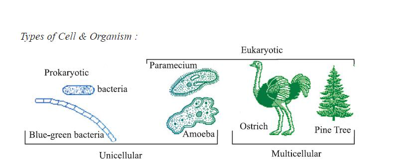
Types of Cells and OrganismsDivision on basis of number of cells
Unicellular
Multicellular
They are composed of single cells
They are composed by multiple cells: usually large number
All functions are performed by the same cell i.e there is no division of labour
Different cells perform specific functions i.e. there is division of labour
Cell division involves only single cell
Specialized cells perform function of reproduction
They usually have shorter lives
Usually have longer lifespan
Examples : Amoeba, Chlamydomonas, Paramoecium and bacteria
Examples : Fungi, plants and animals.
Division on basis of type of organization
Prokaryotic (Primitive Cell)
Eukaryotic
Small in size ; ( 1-10 micrometer)
Comparatively large in size (5-100 micrometer)
Nucleus is not well defined and therefore it is called Nucleoid (nucleus like); not surrounded by nuclear membrane
Well defined nucleus; surrounded by nuclear membrane
Nucleolus is absent
Nucleolus is present
They are uni-cellular
They can be uni-cellular or multi-cellular
Only single chromosome is present
More than one chromosome is present
Cell division is usually by fission or budding
Cell division is by Mitosis or Meiosis
Membrane bound cell organelle are absent
Membrane bound cell organelle are present
Examples : Bacteria, Blue-Green Algae, Mycoplasma
Examples : Plant and Animals
The chlorophyll in photosynthetic prokaryotic bacteria is associated with membranous vesicles (bag like structures) but not with plastids as in eukaryotic cells
Why do some cells have specific shapes and sizes?
The shapes and size of cells depends upon the function they perform.
Some cells should change shape such as Amoeba which changes it shape to perform various functions such as locomotion (movement), ingestion and excretion.
Some cells on the other hand have definite shape as they perform specific functions.
For Example:
-Nerve Cell is elongated to transmit messages
-RBCs are discoidal (disc shaped) to carry blood
-Muscle cells are spindle shaped to provide elasticity and strength.
The same applies to tissues which are made up of specific type of cells and their structure too is dependent upon the kind of function they perform.
In humans, the stem-cell is responsible for generation of all other kinds of cell. Stem cell research has allowed scientists to explore the possibilities of tissue and organ regeneration that can open medical frontiers for human health.
Cell Size:
Size of cell varies with the type of organism.
Some are microscopic while some are visible with naked eyes.
Their size may vary from 0.2 mm to 18 cm.
Size of a typical cell in a multi-cellular organism ranges from 20-30 mm.
The largest cell is ostrich egg (15 cm in diameter with shell & 8 cm in diameter without cell).
The longest cell is nerve cell (upto 1 m or more).
Smallest cells so far known are PPLOs e.g., mycoplasma (0.1 mm in diameter).
Human egg is 0.1 mm in diameter.
Division of labour within a cell.
Each cell has certain specific components within it known as cell organelles.
Each kind of cell organelle performs a special function, such as making new material in the cell, clearing up the waste material from the cell and so on.
A cell is able to live and perform all its functions because of these organelles.
These organelles together constitute the basic unit called the cell.
Components and Structural Organization of Cell
There are three features in every cell:
1. Plasma membrane or Cell membrane
2. Nucleus
3. Cytoplasm
Plant cells in addition to cell membrane have a cell wall as well.
Cytoplasm has several cell organelles that help the cell perform its functions.
Plasma Membrane or Cell membrane or Plasma lemma
It is the outermost covering of the cell that separates the contents of the cell from its
It is found in both, plant and animal cells and is the outermost covering in case of animals and is found below the cell wall in plants.
It is flexible and made up of organic molecules called lipids and proteins but the structure can only be seen through an electron microscope. The proteins are sandwiched between lipids.
The flexibility of the membrane allows some cells to engulf food and other materials from its external environment by a process known as endocytosis as in case of Amoeba.
It separates the cytoplasm from its surroundings by acting as a limiting boundary.
It is selectively permeable, which means that it allows or ‘permits’ the entry and exit of some materials from in and out of the cell.
Functions of Plasma Membrane
It regulates the movement of molecules inside and outside the cell.
It helps in maintaining the distinct composition of the cell.
How does the movement of material take place across cell membrane?
Movement of substances through cell membrane can happen either through Diffusion or Osmosis.
What is Diffusion?
The spontaneous movement of substances (solute or solvent) from a region of high concentration of low concentration is called as diffusion.
It does not require any extra energy and is therefore spontaneous. It can also be called a passive transport.
The transport of material happens in the medium and the presence of a selective membrane is not necessary.
The movement happens till equilibrium concentration is achieved.
For example, Carbon Dioxide or Oxygen move across the membrane through diffusion. But diffusion of gases can happen without a membrane as well (In case of a smell of hot food)
What is Osmosis?
The movement of solvent from a higher concentration to lower concentration through a selectively permeable membrane till the equilibrium is achieved is called as Osmosis.
Only the solvent particles move and it also happens till equilibrium is achieved.
Osmosis therefore can also be called as diffusion of solvents.
Unicellular freshwater organisms and most plant cells tend to gain water through
osmosis. Absorption of water by plant roots is also an example of osmosis.
What is Endosmosis and Exosmosis?
Movement of particles into the cell is called as Endosmosis.
Movement of particles outside the cell is called as Exosmosis.
Viruses lack any membranes and hence do not show characteristics of life until they enter a living body and use its cell machinery to multiply.
Effect of Types of Solution on Cells
(a) Hypertonic Solution: When the concentration of the solution outside the cell is more than the inside the cell. Due to this the cell looses water and becomes plasmolyzed.
When a living plant cell loses water through osmosis there is shrinkage or contraction of the contents of the cell away from the cell wall.
This phenomenon is known as plasmolysis.
(b) Isotonic Solution: When the concentration of the solution outside the cell is equal to the concentration of cytoplasm of the cell, it is called as isotonic solution.
(c) Hypotonic Solution: When the concentration of the solution outside the cell is lesser than that of cytoplasm of cell. Due to this the cell swells up and bursts.
Cell Wall
It is the outermost covering in plant cells and is absent in animal cells.
It is made up of cellulose and hemi-cellulose Cellulose is a complex substance and provides structural strength to plants.
It is a non living structure which is rigid, strong, thick and porous.
Cell walls permit the cells of plants, fungi and bacteria to withstand very dilute (hypotonic) external media without bursting.
The cell swells, building up pressure against the cell wall. The wall exerts an equal pressure against the swollen cell.
Because of their walls, such cells can withstand much greater changes in the surrounding medium than animal cells.
Functions of Cell Wall:
It provides definite shape to the cell.
It provides strength to the cell.
It is permeable and allows entry of molecules of different sizes.
It has the characteristics of repair and regeneration.
Nucleus
Nucleus was discovered by Robert Brown in 1831 and is the most important cell organelle which directs and controls all its cellular activities.
It plays a crucial part, along with the environment, in determining the way the cell will develop and what form it will exhibit at maturity, by directing the chemical activities of the cell.
It is called therefore called as ‘Headquarter of the cell’ or ‘Control Center of the Cell’.
It has double layered covering called as nuclear membrane which has pores to regulate the movement of materials in and out of the nucleus.
Besides nuclear membrane, nucleus also contains nucleolus and chromatin material and the substance filled inside the nucleus is nucleolus.
Chromosomes or chromatin material consists of DNA (Deoxyribose Nucleic Acid) which stores and transmits hereditary information for the cell to function, grow and reproduce.
Chromatin material is visible as entangled mass of thread like structures. Whenever the cell is about to divide, the chromatin material gets organized into chromosomes.
Chromosomes are visible as rod-shaped structures only when the cell is about to divide.
Functional segments of DNA are called genes. In a cell which is not dividing, this DNA is present as part of chromatin material.
In Eukaryotes, a well-defined nucleus is present while in Prokaryotes, a well-defined nucleus is absent. Prokaryotes contain a primitive nucleus called Nucleoid.
Functions of Nucleus:
It controls all the metabolic activities of the cell and regulates the cell cycle.
It helps in transmission of hereditary characters from parents to off springs.
Cytoplasm
The cytoplasm is the fluid content inside the plasma membrane. It also contains many specialized cell organelles.
Each of these organelles performs a specific function for the cell.
Cytoplasm was discovered by Kolliker in 1862.
It is the site of both biosynthetic and catabolic pathways.
Biosynthetic pathways are those processes which are involved in making of complex molecules within the living cell.
Catabolic pathways are the processes involving breakdown of particles for energy.
It can be divided into two parts :
(i) Cytosol: Aqueous soluble part contains various fibrous proteins forming cytoskeleton.
(ii) Cell organelles: Living part of the cells having definite shape, structure and function bounded by plasma membrane.
Large and complex cells, including cells from multicellular organisms, need a lot of chemical activities to support their complicated structure and function. To keep these activities of different kinds separate from each other, these cells use membrane-bound little structures (or ‘organelles’) within themselves.
Cell Organelles
1. Endoplasmic Reticulum
It is the network of membrane bound tubes and sheets present in the cytoplasm used for manufacturing and transport of proteins, fats and lipids to various places in the cell depending upon the need.
Some of these proteins and lipids help in building the cell membrane. This process is known as membrane biogenesis.
Some other proteins and lipids function as enzymes and hormones.
ER varies greatly in appearance in different cells but it always forms a network system.
What do they normally look like?
They look like long tubules or round or oblong bags (vesicles).
The ER membrane is similar in structure to the plasma membrane.
These are present in all cells except prokaryotes and mammalian erythrocytes.
Functions of ER
They serve as channels for the transport of materials (especially proteins) between various
regions of the cytoplasm or between the cytoplasm and the nucleus.
The ER also functions as a cytoplasmic framework providing a surface for biochemical reactions. For example, in liver cells of vertebrates, Smooth ER helps in removal of many toxins from the body.
Types of Endoplasmic Reticulum.
Rough Endoplasmic Reticulum
Smooth Endoplasmic Reticulum
They are rough due to presence of ribosomes on its surface.
They are smooth as they do not have
ribosomes on its surface.
Ribosomes are responsible for protein
synthesis.
They help in manufacture of fat molecules or lipids and important for cell function.
2. Golgi Apparatus
Golgi Apparatus consists of a system of membrane-bound vesicles (flattened sacs).
These are arranged approximately parallel to each other in stacks called cisterns.
They are connected to membranes of Endoplasmic Reticulum and helps in dispatching of material synthesized near ER inside and outside the cell.
It was discovered by Camilo Golgi.
Functions of Golgi Apparatus
It helps in formation of lipids.
It helps in formation of lysosomes
It helps in melanin synthesis.
Lipids and proteins synthesized in endoplasmic reticulum are packed at Golgi and then transported in form of vesicles.
They provide the site for assembly of new membrane material.
3. Lysosomes
They are membrane bound structure filled with digestive enzymes which are made at rough endoplasmic reticulum (RER).
They acts as a waste removal system keeping the cell clean by digesting any foreign material or worn out cell organelles.
When there is disturbance in the metabolic processes of the cell due to any foreign material then the powerful digestive enzymes in the lysosome breakdown them into simpler substances.
In extreme cases, the lysosome itself bursts releasing the enzymes to digest the entire cell and it is therefore also called as the ‘Suicidal Bags’
4. Mitochondria
They are called as the ‘power house of the cell’ as the glucose is converted into energy at this site in the cell.
They have their own DNA and ribosomes and are therefore able to make their own proteins.
It has 2 membranous structures.
- The outer membranous structure is porous for transportation of material
-The inner membranous structure is highly folded to increase the surface area for ATP production.
ATP stands for Adenosine Tri-Phosphate which is the energy currency of the cell. All the energy consumed is first converted into ATP inside the mitochondria and then used up by the cell.
The body uses energy stored in ATP for making new chemical compounds and for mechanical work.
5. Plastids
They are double membranous and disc like structures found only in plant cells.
They also have their own DNA and ribosomes
There are two types of plastids depending upon the pigment they contain:
(i) Leucoplasts: White or colorless plastids. They are generally found in underground parts of the plant.
Leucoplasts are primarily organelles in which materials such as starch, oils and protein granules are stored.
(ii) Chromoplasts:
These are plastids that contain colored pigments.
Chromoplasts that contain the pigment chlorophyll are known as chloroplasts.
Chloroplasts are important for photosynthesis in plants. Chloroplasts also contain various yellow or orange pigments in addition to chlorophyll.
The internal organization of the Chloroplast consists of numerous membrane layers embedded in a material called the stroma.
These are similar to mitochondria in external structure.
6. Vacuoles
These are storage sacs for liquid or solid contents.
Vacuoles in Plant Cells
In plant cells, there is generally a single large vacuole that can occupy upto 50-90% of the cell space
In plant cells vacuoles are full of cell sap and provide turgidity and rigidity to the cell.
Many substances of importance in the life of the plant cell are stored in vacuoles.
These include amino acids, sugars, various organic acids and some proteins. In
Vacuoles in Animal Cells
In animal cells, there are generally several small sized vacuoles. In Amoeba, the food vacuole
contains the food items that the Amoeba has consumed.
Functions of Vacuoles
-They help in maintaining osmotic pressure of the cell.
-The help in providing rigidity and turgidity.
-The acts as storage units.
-In some cells, they may be used to expel excess water or wastes.
How is the structural organization of the cell important for ensuring its proper functioning?
Each cell acquires its structure and ability to function because of the organization of its membrane and organelles in specific ways.
The cell thus has a basic structural organization.
This helps the cells to perform functions like respiration, obtaining nutrition, and clearing of waste material, or forming new proteins.
Difference between Plant and Animal Cells
Plant Cell
Animal Cell
They have a cell wall to help maintain its shape, turgidity and rigidity along with the cell membrane
Only cell membrane is present.
They contain chloroplasts
They do not contain chloroplasts
They have generally single large vacoule
They have many smaller vacuoles
They have limited movement
They can generally move around
They do not have centrioles
They have centrioles
Nucleus is generally on the side
Nucleus is generally in the center
Key takeaways
The cell is the fundamental structural and functional unit of all living organisms, first identified by Robert Hooke in 1665.
The Cell Theory establishes that all life is composed of cells, and all new cells arise from pre-existing cells.
Cells are bounded by a selectively permeable plasma membrane that regulates the movement of substances via diffusion and osmosis.
Eukaryotic cells contain specialized membrane-bound organelles, such as mitochondria for energy production and lysosomes for waste disposal.
Plant cells differ from animal cells primarily by the presence of a rigid cellulose cell wall, chloroplasts for photosynthesis, and large central vacuoles.
Test yourself
Who coined the term 'cell' and what was he observing?
Robert Hooke coined the term in 1665 while observing a thin slice of cork under a primitive microscope.
What is the primary function of the mitochondria?
Mitochondria act as the powerhouses of the cell, generating energy in the form of ATP molecules.
How does a prokaryotic cell differ from a eukaryotic cell regarding its nucleus?
A prokaryotic cell lacks a membrane-bound nucleus, storing its DNA in an undefined nucleoid region, whereas a eukaryotic cell has a true, membrane-bound nucleus.
Play with the idea
The fundamental unit of life: test the theory, match the organelle, predict the transport
These scenarios explore cell theory evidence, prokaryote vs eukaryote distinctions, organelle structure-function relationships, membrane transport mechanisms, and tonicity effects. Each question uses fictional but structurally accurate experimental evidence. The test is whether you can connect the evidence to the correct cellular explanation.
Situation 1
Explore the reasoning for every approach
Correct — Schleiden and Schwann established the cell theory including cell origin.
Incorrect. Schleiden and Schwann established the first two tenets: (1) all organisms are made of cells, (2) the cell is the basic unit of life. They did NOT establish the third tenet (cells from pre-existing cells) — they believed in free-cell formation (crystallisation). Virchow (1855) provided the third tenet based on cell division observations.
Incorrect — Schleiden and Schwann did not address cell origin; Virchow added the third tenet later.
Yes. The modern cell theory has three tenets. Tenet 1 (all living things are cells) and Tenet 2 (cell = basic unit) came from Schleiden (plants) and Schwann (animals) in 1838–39. Tenet 3 (cells from cells) came from Virchow in 1855. The student conflates the contributors.
Partially correct — Schleiden and Schwann proved it for plants and animals respectively.
The issue is not plant vs animal — it's the mechanism of cell origin. Both Schleiden and Schwann thought new cells formed spontaneously from a "cytoblastema" (crystallisation). They did not observe cell division as the source. Virchow's contribution was showing division is the only source.
Situation 2
Explore the reasoning for every approach
Correct — plant cells have cell walls, so this must be a plant cell.
Incorrect. The cell wall composition is the key: peptidoglycan is unique to bacteria (prokaryotes). Plant cell walls are cellulose. Additionally, circular DNA in a nucleoid (no nuclear membrane), 70S ribosomes, and absence of mitochondria are all prokaryotic features. This is a bacterium, not a plant cell.
Incorrect — peptidoglycan wall + circular DNA + 70S ribosomes = prokaryote (bacterium), not plant.
Yes. The evidence checklist: (1) Nucleoid (no nucleus) = prokaryote. (2) Circular DNA = prokaryote. (3) 70S ribosomes = prokaryote (eukaryotes have 80S). (4) No mitochondria = prokaryote. (5) Peptidoglycan cell wall = bacteria specifically. All five point to a bacterial cell. Plant cells are eukaryotic: nucleus, linear DNA, 80S ribosomes, mitochondria, cellulose wall.
Cannot determine — some bacteria have cellulose walls too.
No bacteria have cellulose walls. Cellulose is unique to plants and some algae. Peptidoglycan is the defining bacterial cell wall polymer. The features are mutually exclusive between prokaryotes and eukaryotes.
Situation 3
Explore the reasoning for every approach
Correct — the wall blocks water, so the plant cell doesn't swell.
Incorrect. The plant cell DOES take in water (it becomes turgid). The wall does not block water entry — it is freely permeable. The wall exerts an opposing pressure (wall pressure) that balances the osmotic pressure, preventing lysis. Water still enters until pressure equilibrium. The animal cell has no wall, so pressure cannot build, and it bursts.
Incorrect — water enters both cells; the plant wall provides pressure resistance, not a water barrier.
Yes. Osmosis occurs in both cells (water moves into both). The difference is mechanical: the plant cell wall is rigid and pushes back (turgor pressure), reaching equilibrium without bursting. The animal cell membrane cannot resist, so it stretches until it ruptures. The wall is not a water barrier — it is a pressure vessel.
Partially correct — the wall slows water entry enough to prevent bursting.
The wall does not slow water entry significantly. Water crosses the cell membrane (same in both) at similar rates. The turgid plant cell has taken up as much water as the animal cell before lysis; the difference is the wall's structural resistance, not permeability.
Investigate before you memorise
The fundamental unit of life: cells, organelles, and how they exchange with the world
Explore the evidence that built cell theory, the architecture of prokaryotes vs eukaryotes, and the transport mechanisms that keep cells alive. Make a prediction before opening each section.
Open the cell theory timeline
From "small rooms" to "all cells from cells" — 200 years of evidence
Each scientist added a piece. The modern theory has three tenets: (1) All living things are made of cells. (2) The cell is the basic unit of life. (3) All cells arise from pre-existing cells.
Scientist
Contribution
Limitation
Robert Hooke (1665)
Observed cork cells; coined "cell" (Latin cellula = small room)
Saw only dead cell walls; no living content
Antonie van Leeuwenhoek (1674)
First to observe living cells (bacteria, protozoa, spermatozoa) with improved microscope
No systematic theory; called them "animalcules"
Robert Brown (1831)
Discovered nucleus in orchid cells
Did not generalise to all cells
Matthias Schleiden (1838)
All plants are made of cells
Plant cells only; thought cells formed by crystallisation
Theodor Schwann (1839)
All animals are made of cells; unified plant + animal cell theory
Still thought cells formed spontaneously
Rudolf Virchow (1855)
"Omnis cellula e cellula" — all cells arise from pre-existing cells
Completed the theory; no major limitation
Why is Virchow's "Omnis cellula e cellula" the final piece?
Schleiden and Schwann established that plants and animals are made of cells, but they thought new cells formed by crystallisation (free-cell formation). Virchow showed cells only come from division of existing cells — this explained growth, repair, and reproduction, and refuted spontaneous generation at the cellular level. The three tenets together define the cell as the unit of continuity of life.
Open the prokaryote vs. eukaryote comparison
The great divide: nucleus or no nucleus
Every cellular feature follows from this one difference. Eukaryotes compartmentalise; prokaryotes don't.
Feature
Prokaryote
Eukaryote
Size
1–10 µm
10–100 µm
Nucleus
Absent (nucleoid region)
Present (membrane-bound)
Membrane-bound organelles
Absent
Present (mitochondria, ER, Golgi, lysosomes...)
DNA
Circular, single chromosome, no histones
Linear, multiple chromosomes, with histones
Ribosomes
70S (50S + 30S)
80S (60S + 40S); also 70S in mitochondria/chloroplasts
Why do eukaryotes have 80S ribosomes but mitochondria have 70S?
Mitochondria evolved from an ancient aerobic bacterium (endosymbiosis). They retained their own 70S ribosomes, circular DNA, and double membrane. The host cell's cytoplasmic ribosomes are 80S. This is evidence for endosymbiotic theory — also true for chloroplasts. Antibiotics targeting 70S ribosomes affect bacteria and mitochondria (side effects), but not cytoplasmic 80S ribosomes.
Open the organelle atlas
Ten organelles — structure matches function
Each organelle is a specialised compartment. The membrane system (nucleus, ER, Golgi, vesicles, lysosomes, vacuole, cell membrane) works as an integrated factory.
Organelle
Structure
Function
Found In
Cell membrane
Phospholipid bilayer with embedded proteins (fluid mosaic)
RNA + protein; 70S (prokaryotes) or 80S (eukaryotes)
Protein synthesis (translation); free or ER-bound
Both
Why is the nucleus not in the "membrane system" trafficking pathway?
The nucleus communicates via nuclear pores (import/export of RNA, proteins), not vesicles. The ER, Golgi, lysosomes, vacuole, and cell membrane are connected by vesicular transport. The nuclear envelope is continuous with rough ER, but nuclear transport is pore-mediated, not vesicular. This distinction matters for understanding protein targeting signals.
Open the transport mechanism lab
Six ways across the membrane — only two use ATP directly
Passive = down gradient, no ATP. Active = against gradient, uses ATP. Bulk transport = vesicles.
Type
Energy
Direction
Examples
Diffusion
Passive (no ATP)
High → low concentration
O₂, CO₂, water (osmosis), small nonpolar molecules
Facilitated diffusion
Passive (no ATP)
High → low concentration
Glucose, amino acids, ions via channels/carriers
Osmosis
Passive (no ATP)
Water: low solute → high solute
Water across semipermeable membrane
Active transport
Active (uses ATP)
Low → high concentration
Na⁺/K⁺ pump, proton pump, nutrient uptake against gradient
Why does facilitated diffusion need a protein but not ATP?
The protein (channel or carrier) provides a hydrophilic pathway for polar/charged molecules that cannot cross the lipid bilayer. The driving force is still the concentration gradient — the protein just removes the barrier. No ATP is used because the movement is downhill. Contrast with active transport: the protein (pump) uses ATP to move against the gradient.
Open the tonicity game
Predict the cell's fate in each solution
Water follows solute. The cell wall changes the outcome for plants.
Solution
Water Movement
Animal Cell
Plant Cell
Isotonic
No net movement
Normal shape
Normal (flaccid)
Hypotonic
Into cell
Swells, may burst (lysis)
Swells, turgid (wall prevents bursting)
Hypertonic
Out of cell
Shrinks (crenation)
Plasmolysis (membrane pulls from wall)
Why don't plant cells burst in hypotonic solution?
The rigid cell wall (cellulose) exerts an opposing pressure (wall pressure) that balances the inward osmotic pressure. The cell becomes turgid — this is the healthy state for plants. Animal cells lack a wall, so uncontrolled water entry causes lysis. This is why IV fluids must be isotonic (0.9% saline).
Open the identification game
Identify the organelle, the transport, or the cell type
Q: Organelle with own DNA, double membrane, cristae. Prediction: Mitochondria.
Q: Protein-studded membrane sack that modifies and sorts. Prediction: Golgi apparatus.
Q: Glucose enters a cell via a carrier protein, down its gradient. Prediction: Facilitated diffusion.
Q: Na⁺ pumped out, K⁺ pumped in, using ATP. Prediction: Active transport (Na⁺/K⁺ pump).
Q: Cell placed in solution; swells and becomes turgid. Prediction: Plant cell in hypotonic solution.
Q: Circular DNA, 70S ribosomes, no nucleus. Prediction: Prokaryote (bacterium).
Why are lysosomes called "suicide bags"?
If a lysosome membrane ruptures, its hydrolytic enzymes (active at pH ~5) are released into the cytoplasm (pH ~7.2). They digest cellular components, leading to cell death. This is a controlled mechanism for removing damaged cells. In apoptosis, lysosomes are deliberately triggered to release enzymes.
Test your understanding of the cell theory and cellular structure.
1Rudolf Virchow’s 1855 contribution to the Cell Theory was to propose that:
Based on the text, what was Virchow’s key insight and why was it significant?
This was actually the conclusion of Schleiden and Schwann in the 1830s. Their work established that both plants and animals are composed of cells.
While this is a pillar of modern cell theory, Virchow’s specific contribution was different. He focused on the origin of cells, not their structural role.
Virchow stated 'Omnis cellula e cellula' (all cells arise from pre-existing cells). This was significant because it refuted the idea of spontaneous generation and established that cell division is how life continues.
This describes the composition of chromosomes, which the text assigns to the nucleus. This was not Virchow’s contribution to cell theory.
2The text describes the plasma membrane as a "highly sophisticated security gate" that is selectively permeable.
If a plant cell is placed in a salt solution with higher concentration than the cell’s contents, what would most likely happen based on the text’s explanation?
While oxygen does diffuse through the membrane, this would not be the primary outcome. The text emphasizes that osmosis specifically describes water movement across membranes.
The text defines osmosis as 'the diffusion of water across a membrane.' Since the external solution is hypertonic (higher solute concentration), water would move out of the cell to balance concentrations, causing the cell to shrink.
Lysosomes bursting (releasing 'suicide bags') happens when a cell is damaged, not due to external salt concentration. This is unrelated to the membrane's selective permeability.
DNA is contained in the nucleus (as chromatin or chromosomes) and cannot simply pass through the plasma membrane. The membrane’s selective permeability actually prevents such large molecules from moving freely.