
Nobel Prize in Physiology or Medicine 2003: Magnetic Resonance Imaging and Its Discoverers
On this page
What was the Nobel Prize in Physiology or Medicine 2003 awarded for?
The Nobel Assembly at Karolinska Institutet awarded the prize jointly to two scientists "for their discoveries concerning magnetic resonance imaging". This is the official citation, and it is worth reading slowly because every word matters.
"Magnetic resonance imaging" (MRI) is a method of making pictures of the inside of the human body without cutting it open and without using harmful radiation.
The laureates did not invent the underlying physics of magnetic resonance, which had already won a Nobel Prize in Physics in 1952.
Instead, in the early 1970s, they worked out how to turn that physics into a practical imaging technique that could show soft tissues such as the brain, spinal cord and internal organs in detail.
The prize itself is formally called the Nobel Prize in Physiology or Medicine. It is one of the original five prizes set out in Alfred Nobel's will and is awarded each year by the Nobel Assembly at Karolinska Institutet in Sweden, usually announced in early October and presented in December.
Who are the laureates?
The 2003 prize was shared equally between a chemist working in the United States and a physicist working in the United Kingdom, reflecting how the discovery grew from basic physics and chemistry into a medical tool.
Paul C. Lauterbur
Paul C. Lauterbur was born on 6 May 1929 in Sidney, Ohio, USA, and died on 27 March 2007 in Urbana, Illinois, USA. At the time of the award he was affiliated with the University of Illinois, Urbana, Illinois, USA.
He received one half of the prize. In the early 1970s, Lauterbur discovered that introducing variations, or gradients, into the magnetic field used in magnetic resonance made it possible to build a two-dimensional picture.
By analysing the characteristics of the radio waves given off by the sample, he could work out exactly where in space each signal had come from.
In 1973 he described how adding gradient magnets to the main magnetic field let him visualise a cross-section of tubes filled with ordinary water surrounded by heavy water, something no other technique can distinguish.
Sir Peter Mansfield
Sir Peter Mansfield was born on 9 October 1933 in London, United Kingdom, and died on 8 February 2017. At the time of the award he worked at the University of Nottingham, School of Physics and Astronomy, United Kingdom.
He also received one half of the prize. Mansfield developed the mathematical methods needed to analyse the gradient signals quickly and turn them into a usable image, which made the technique practical rather than merely possible.
He further showed that it was feasible, in principle, to capture images extremely fast using rapid variations in the magnetic-field gradients, an approach now called echo-planar imaging. This fast-scanning idea became clinically useful roughly a decade after he proposed it.
What problem were the laureates trying to solve?
Doctors have always needed ways to look inside the living body without cutting it open.
By the mid-twentieth century, X-rays and later computer tomography (CT, itself awarded a Nobel Prize in Physiology or Medicine in 1979) allowed internal imaging, but both rely on ionizing radiation, which carries some risk with repeated use and does not show soft tissue contrast especially well.
Meanwhile, physicists had already discovered the phenomenon behind MRI. In 1946, Felix Bloch and Edward Mills Purcell in the United States demonstrated nuclear magnetic resonance (NMR): atomic nuclei placed in a magnetic field absorb and then re-emit radio waves at a frequency that depends on the field strength.
This earned them the Nobel Prize in Physics in 1952. For the following decades, NMR was used mainly by chemists to study the structure of substances in a laboratory sample, not to image a whole human body.
The gap that remained was this: NMR signals from a sample told scientists about the chemistry present, but not where in space each part of the signal came from.
Without that spatial information, there was no way to build a picture. Solving this gap, in the early 1970s, is exactly what Lauterbur and Mansfield each contributed to in different ways, turning a chemistry tool into a medical imaging method.
How does magnetic resonance work, from the ground up?
To understand MRI, start with a single hydrogen atom. Its nucleus is a single proton, and protons behave like tiny spinning magnets, each with its own small magnetic moment.
The human body is useful here because roughly two-thirds of body weight is water, and water molecules each contain two hydrogen atoms, so the body is rich in these tiny magnetic nuclei.
When a person is placed inside a strong, steady magnetic field, these hydrogen nuclei line up, loosely, along the direction of the field, rather like compass needles all pointing roughly the same way. Left alone, they would simply sit there.
But if the body is then hit with a pulse of radio waves at exactly the right frequency, the nuclei absorb energy and their alignment is disturbed; this matching of frequencies is called resonance.
After the radio pulse ends, the disturbed nuclei gradually return to their original alignment, and as they do, they give off their own radio-wave signal.
Different tissues contain different amounts of water, and water in a diseased tissue often behaves slightly differently from water in healthy tissue, so the strength and timing of the returning signal varies from place to place.
This variation is the raw information that a scanner has to turn into a picture.
How did Lauterbur and Mansfield turn resonance into an image?
Having a radio signal from the whole body is not enough; a doctor needs to know where exactly each part of the signal came from. This spatial problem is what the two laureates solved, using slightly different but complementary methods.
- A strong, uniform magnetic field is applied across the body so that hydrogen nuclei align in a known, predictable way.
- A smaller, deliberately uneven magnetic field, called a gradient, is added on top of the main field, so that the overall field strength (and therefore the resonance frequency) differs slightly from one point in the body to another.
- A pulse of radio waves is sent in, tuned to the resonance frequencies now present across the gradient; the nuclei at each position absorb and later emit signals at a slightly different frequency depending on exactly where they are.
- The returning radio signals are picked up and their frequencies are analysed; since each frequency corresponds to a known position in the gradient, this analysis reveals where each part of the signal originated, the step Lauterbur pioneered.
- The signals are then processed mathematically, and rapidly, to reconstruct them into a coherent image, the computational step Mansfield developed and later extended into very fast scanning.
- The processed signal strengths at each point are assembled into a two- or three-dimensional picture, where brightness reflects differences in water content and behaviour between tissues.
Diagram
Lauterbur's gradient-imaging experiment
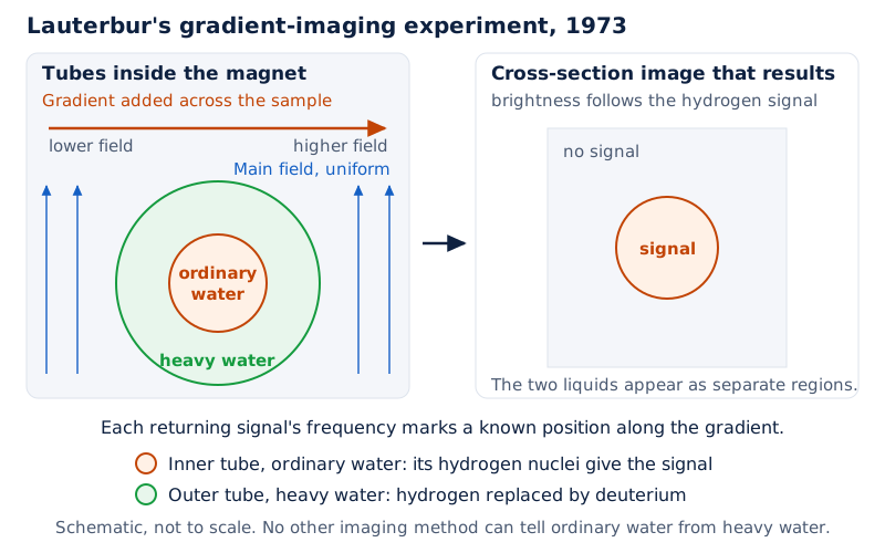
Sketch a tube of ordinary water sitting inside a larger tube of heavy water, both placed in a magnetic field with a gradient running across them.
Label the gradient direction, and show how the two liquids, indistinguishable to other methods, appear as separate regions once gradient-based imaging is applied, since only ordinary water contains the hydrogen nuclei being detected.
Drawn by One Young India.
This is why the presentation speech compared plain NMR spectroscopy to hearing a symphony on an old radio, and imaging with gradients to sitting in the concert hall and actually seeing which instrument is playing which note; a false note, in medicine, is a disease process in the body.
What does MRI reveal in the body?
Because the signal depends on water content and on how water molecules move and interact with surrounding tissue, MRI is particularly sensitive to changes that other imaging methods miss.
A difference in water content of less than one percent between healthy and diseased tissue can already show up on an MRI picture. This sensitivity has made MRI especially valuable in certain parts of medicine, summarised below.
| Clinical use | What MRI shows or replaces |
|---|---|
| Multiple sclerosis | Location, intensity and treatment response of inflammation in the brain and spinal cord |
| Lower back pain | Whether a disc herniation is pressing on a nerve or the spinal cord, guiding the need for surgery |
| Brain and movement-disorder surgery | Precise placement of electrodes in central brain nuclei for pain or Parkinson's disease treatment |
| Cancer diagnosis and staging | Exact tumour limits, depth of infiltration (for example in colon cancer) and lymph node involvement |
| Pancreatic and bile duct examination | Replaces an invasive endoscope-and-contrast procedure that can cause complications |
| Knee joint examination | Detailed views of cartilage and cruciate ligaments, replacing invasive arthroscopy |
In every one of these uses, the underlying signal is the same one Lauterbur and Mansfield learned to locate and reconstruct: hydrogen nuclei in water, responding to a magnetic field and radio pulses.
How did the discovery unfold?
The route from a basic physics observation to a routine hospital scanner took several decades and involved more than one Nobel Prize, which helps explain why the prize went to two people working independently on complementary pieces of the puzzle.
| Year | Event |
|---|---|
| 1946 | Felix Bloch and Edward Mills Purcell demonstrate nuclear magnetic resonance in protons |
| 1952 | Bloch and Purcell are awarded the Nobel Prize in Physics for this discovery |
| Early 1970s | Paul Lauterbur discovers that adding gradients to the magnetic field allows a two-dimensional image to be built |
| 1973 | Lauterbur describes imaging a cross-section of ordinary water surrounded by heavy water using gradient magnets |
| 1970s | Peter Mansfield develops the mathematical analysis of gradient signals and proposes extremely fast echo-planar imaging |
| Early 1980s | The first MRI equipment becomes available for use in hospitals |
| 1991 | Richard Ernst is awarded the Nobel Prize in Chemistry for high-resolution NMR spectroscopy methodology |
| 2002 | About 22,000 MRI machines are in use worldwide, performing over 60 million examinations a year; Kurt Wüthrich is awarded the Nobel Prize in Chemistry for NMR structure determination of biological macromolecules |
| 2003 | Paul C. Lauterbur and Sir Peter Mansfield are jointly awarded the Nobel Prize in Physiology or Medicine |
Why does it matter?
MRI is now a routine diagnostic method used on almost every organ of the body, and the Nobel committee noted that the method was still developing rapidly even at the time of the award.
Its biggest practical advantage, according to the press release, is that it is harmless according to all present knowledge, because unlike X-ray or CT it does not use ionizing radiation.
There are limits. Patients who have magnetic metal implants or a pacemaker cannot safely be scanned because of the strong magnetic field, and patients with claustrophobia can find the enclosed scanner difficult.
Even so, MRI has replaced several more invasive and uncomfortable examinations, reducing risk and discomfort for many patients, for example by avoiding contrast-injection endoscopy of the bile ducts or diagnostic arthroscopy of the knee.
The committee also stressed that MRI contributes across the whole chain of healthcare, from screening and detection through diagnosis and treatment planning to following up how a disease responds over time, which is why the discovery was judged to have had such broad medical importance.
How does this connect to what you study?
The physics behind MRI connects directly to topics on atomic structure and magnetism studied in school science: protons behaving as tiny magnets, the idea of resonance between a wave and a system tuned to the same frequency, and the difference between ionizing radiation (X-rays) and non-ionizing methods.
It also links to basic biology: the human body is roughly two-thirds water, hydrogen atoms are abundant because of this water content, and different tissues and disease states hold water differently, which is exactly what a scanner is designed to detect.
Seeing how a physics discovery from 1946 fed into a medical breakthrough by the 1970s, and into routine hospital use by the 1980s, is also a useful real example of how long basic research can take to reach patients.
Quick facts for exams
The Nobel Prize in Physiology or Medicine 2003 was awarded jointly to Paul C. Lauterbur of the University of Illinois, USA, and Sir Peter Mansfield of the University of Nottingham, United Kingdom, each receiving one half of the prize.
The citation reads "for their discoveries concerning magnetic resonance imaging". The prize was announced on 6 October 2003 by the Nobel Assembly at Karolinska Institutet. Lauterbur discovered that gradients in a magnetic field allow two-dimensional imaging;
Mansfield developed the mathematics to analyse gradient signals and showed very fast imaging was possible.
Their work, building on the 1946 discovery of nuclear magnetic resonance by Bloch and Purcell, led to modern MRI, now used worldwide for diagnosing diseases of the brain, spine and other organs without ionizing radiation.
| Fact | Detail |
|---|---|
| Prize | Nobel Prize in Physiology or Medicine 2003 |
| Laureates | Paul C. Lauterbur and Sir Peter Mansfield |
| Country of birth | Lauterbur: USA (Sidney, Ohio); Mansfield: United Kingdom (London) |
| Affiliation at award | Lauterbur: University of Illinois, Urbana, USA; Mansfield: University of Nottingham, UK |
| Shares | One half each |
| Citation | "for their discoveries concerning magnetic resonance imaging" |
| Date announced | 6 October 2003 |
| Prize amount | 10,000,000 Swedish kronor |
Note: Source. The prize facts in this note are from the Nobel Prize's official site, nobelprize.org.
Glossary
- Magnetic resonance imaging (MRI) — a method that uses magnetic fields and radio waves to make detailed pictures of soft tissue inside the body without surgery or ionizing radiation.
- Nuclear magnetic resonance (NMR) — the physical phenomenon in which atomic nuclei in a magnetic field absorb and re-emit radio waves at a frequency matched to the field strength.
- Resonance — the matching of a radio-wave frequency to the natural frequency of a spinning nucleus, allowing energy to be absorbed.
- Gradient (magnetic field gradient) — a deliberately uneven magnetic field, added to the main field, that makes resonance frequency depend on position.
- Proton — a positively charged particle in an atomic nucleus; the hydrogen nucleus consists of a single proton, which behaves like a tiny magnet.
- Echo-planar imaging — a very fast MRI scanning method, proposed by Mansfield, using rapid changes in the gradient field.
- Ionizing radiation — high-energy radiation, as used in X-rays and CT, that can damage tissue; MRI does not use it.
- Computer tomography (CT) — an earlier X-ray-based imaging method, itself awarded the Nobel Prize in Physiology or Medicine in 1979.
- Spectroscopy — the earlier use of magnetic resonance to study chemical structure in a laboratory sample, before imaging was developed.
- Heavy water — water in which the hydrogen is replaced by deuterium, used by Lauterbur to show that MRI could distinguish it from ordinary water.
- Multiple sclerosis — a disease causing inflammation in the brain and spinal cord, for which MRI is especially useful in diagnosis and follow-up.
- Disc herniation — a condition where a spinal disc presses on a nerve, which MRI can detect without invasive testing.
Common errors and misconceptions
- Misconception: Lauterbur and Mansfield discovered nuclear magnetic resonance itself. Correct: NMR was discovered by Bloch and Purcell in 1946 and earned the 1952 Nobel Prize in Physics; the 2003 laureates discovered how to turn NMR into an imaging method.
- Misconception: MRI uses X-rays or radioactive material. Correct: MRI uses magnetic fields and radio waves and, according to the committee, does not use ionizing radiation at all.
- Misconception: The prize was for inventing the MRI scanner as a finished machine. Correct: The citation is for discoveries concerning magnetic resonance imaging, the scientific principles that made imaging possible, developed in the early 1970s.
- Misconception: Only one person is credited with inventing MRI. Correct: The prize was shared equally between Lauterbur, who solved spatial localisation, and Mansfield, who solved fast signal analysis.
- Misconception: MRI can be used on every patient. Correct: Patients with magnetic metal implants or pacemakers cannot safely be scanned, and claustrophobic patients may struggle with the enclosed scanner.
- Misconception: MRI detects bone best. Correct: MRI is especially valuable for soft tissue such as the brain, spinal cord and joint cartilage, since it relies on water content differences.
- Misconception: Richard Ernst and Kurt Wüthrich shared this medicine prize. Correct: They won separate Nobel Prizes in Chemistry, in 1991 and 2002 respectively, for related but distinct NMR work.
Exam-style questions with model answers
Q1. What is the official citation for the Nobel Prize in Physiology or Medicine 2003? [2 marks]
- The citation reads "for their discoveries concerning magnetic resonance imaging".
- It was awarded jointly to Paul C. Lauterbur and Sir Peter Mansfield.
Q2. In which year was nuclear magnetic resonance first demonstrated, and by whom? [2 marks]
- Nuclear magnetic resonance was first demonstrated in 1946 by Felix Bloch and Edward Mills Purcell.
- They later won the 1952 Nobel Prize in Physics for this discovery.
Q3. Name the two laureates of the 2003 prize and their affiliations at the time of the award. [2 marks]
- Paul C. Lauterbur was at the University of Illinois, USA.
- Sir Peter Mansfield was at the University of Nottingham, United Kingdom.
Q4. Explain what a "gradient" in the magnetic field does and why it was essential to creating an MRI picture. [4 marks]
- A gradient is a deliberately uneven magnetic field added on top of the strong, uniform main field used in MRI.
- Because resonance frequency depends on local field strength, the gradient makes nuclei at different positions resonate at slightly different frequencies.
- By measuring the frequency of the returning radio signal, Lauterbur could work out exactly where in the body each part of the signal had come from.
- Without a gradient, a scanner would only know that a signal existed, not where it originated, so no spatial image could be built.
Q5. Describe the contribution of Sir Peter Mansfield to magnetic resonance imaging. [4 marks]
- Mansfield further developed the use of gradients in the magnetic field to more precisely distinguish differences in resonance signals.
- He worked out how the detected radio signals could be mathematically analysed quickly and transformed into a usable image.
- This mathematical step was essential in turning gradient imaging from a research curiosity into a practical clinical method.
- He also showed that extremely rapid imaging, later called echo-planar imaging, was achievable using fast gradient variations, which became useful in clinics about a decade later.
Q6. Why is water content important to how MRI produces images of the body? [4 marks]
- About two-thirds of human body weight is water, and each water molecule contains two hydrogen atoms whose nuclei act like tiny spinning magnets.
- In a strong magnetic field these hydrogen nuclei align, and radio pulses at the resonance frequency disturb that alignment.
- Different tissues and organs have different water content, and diseased tissue often shows altered water content compared to healthy tissue.
- These differences change the strength and timing of the returning radio signal, which the scanner converts into brightness differences in the final image, revealing pathological changes.
Q7. Discuss why MRI has become important in medical diagnosis, with examples. [6 marks]
- MRI is a non-invasive method that does not use ionizing radiation, unlike X-rays or computer tomography, making it harmless according to present knowledge for most patients.
- It is especially valuable for imaging the brain and spinal cord, since a water content change of less than one percent can reveal a pathological change.
- In multiple sclerosis, MRI shows exactly where inflammation is located in the nervous system, how intense it is, and how it responds to treatment.
- For lower back pain, MRI can show whether a disc herniation is pressing on a nerve, helping doctors decide if surgery is necessary.
- In cancer, MRI reveals the precise limits and depth of a tumour and whether lymph nodes are affected, improving surgical and radiation planning.
- MRI has also replaced several invasive procedures, such as contrast-injection endoscopy of the bile ducts and diagnostic knee arthroscopy, reducing risk and discomfort for patients.
Q8. Trace, with years, how the discovery of nuclear magnetic resonance led to the clinical use of MRI. [6 marks]
- In 1946, Felix Bloch and Edward Mills Purcell demonstrated nuclear magnetic resonance in protons, work recognised with the 1952 Nobel Prize in Physics.
- For the following decades, scientists used magnetic resonance mainly for spectroscopy, studying the chemical structure of substances rather than imaging bodies.
- In the early 1970s, Paul Lauterbur discovered that adding gradients to the magnetic field allowed a two-dimensional image to be built, and in 1973 he demonstrated this by imaging ordinary water surrounded by heavy water.
- Around the same period, Peter Mansfield worked out the mathematics to analyse gradient signals quickly and proposed that extremely fast imaging was achievable.
- The first MRI equipment became available in hospitals in the early 1980s, roughly a decade after the key discoveries, once Lauterbur's gradient method and Mansfield's mathematical analysis had been engineered into practical scanners.
- By 2002, around 22,000 MRI machines were operating worldwide performing over 60 million scans a year, leading to the joint award of the 2003 Nobel Prize in Physiology or Medicine to Lauterbur and Mansfield.
Key takeaways
- The 2003 Nobel Prize in Physiology or Medicine went jointly to Paul C. Lauterbur and Sir Peter Mansfield for discoveries concerning magnetic resonance imaging.
- Lauterbur discovered that adding gradients to a magnetic field allows a two-dimensional image to be built from resonance signals.
- Mansfield developed the mathematics to analyse gradient signals fast and proposed extremely rapid echo-planar imaging.
- MRI relies on hydrogen nuclei in water behaving like tiny magnets that resonate with radio waves in a magnetic field.
- Nuclear magnetic resonance itself was discovered earlier, in 1946, by Bloch and Purcell, who won the 1952 Nobel Prize in Physics.
- MRI does not use ionizing radiation, unlike X-ray or CT, making it a comparatively safe imaging method.
- MRI is especially valuable for the brain, spinal cord, cancer staging and knee joint examination, and has replaced several invasive procedures.
- By 2002, roughly 22,000 MRI machines performed over 60 million examinations worldwide each year.
Test yourself
Who shared the Nobel Prize in Physiology or Medicine 2003, and in what proportion?
Paul C. Lauterbur and Sir Peter Mansfield shared the prize equally, each receiving one half, for their discoveries concerning magnetic resonance imaging.
What physical property of protons makes MRI possible?
Protons behave like tiny spinning magnets, so they align in a magnetic field and can absorb and re-emit radio waves at a matching resonance frequency.
Why did Lauterbur need to add gradients to the magnetic field?
Gradients make the resonance frequency depend on position, so analysing the returning signal's frequency reveals exactly where in the body it came from.
What did Peter Mansfield contribute beyond gradient imaging itself?
Sir Peter Mansfield worked out how to mathematically analyse gradient signals quickly and showed that extremely fast imaging, called echo-planar imaging, was achievable.
Why is MRI considered safer than X-ray or CT scanning?
MRI uses magnetic fields and radio waves rather than ionizing radiation, so it is harmless according to all present knowledge, unlike X-ray or CT.
Which earlier Nobel Prize recognised the basic phenomenon behind MRI, and to whom?
Felix Bloch and Edward Mills Purcell won the 1952 Nobel Prize in Physics for discovering nuclear magnetic resonance in 1946.
Give one disease where MRI is described as especially valuable for diagnosis and follow-up.
MRI is especially valuable for multiple sclerosis, showing the location, intensity and treatment response of inflammation in the brain and spinal cord.
Why can some patients not undergo an MRI scan?
Patients with magnetic metal implants or a pacemaker cannot be scanned safely because of the scanner's strong magnetic field, and claustrophobic patients may find it difficult.