
The structure of a tooth, different types of teeth | ICSE Class 9 Biology Notes
On this page
This note covers chewing, the external and internal structure of a human tooth, the shapes and functions of different teeth, milk and permanent teeth, the adult dental formula, labelled tooth diagrams, and the development of tooth decay.
How do teeth help to prepare food in the mouth?
Teeth are hard structures in the mouth that cut, tear and grind food. Mastication means chewing. During chewing, teeth break food mechanically into smaller pieces. Mechanical breakdown changes the size of the pieces; it does not itself convert a food substance into a different substance.
The buccal cavity is the cavity of the mouth. Food enters this cavity during ingestion, which means taking food into the body. Teeth begin its physical breakdown here, while the tongue moves the food and mixes it with liquid.
How do teeth, saliva and the tongue work together?
Saliva is the fluid released into the mouth by the salivary glands, the organs that produce it. Saliva wets food, helping its passage through the digestive tract. The tongue is a muscular organ that moves food around during chewing and helps in swallowing.
- Food is taken into the mouth by ingestion, bringing it into contact with the teeth.
- The teeth break the food mechanically into small pieces as it is chewed.
- The tongue moves the food around and mixes it thoroughly with saliva during chewing.
- The tongue helps the chewed food to be swallowed and passed into the foodpipe, the tube leading from the mouth towards the stomach.
The foodpipe, also called the oesophagus, is the tube through which swallowed food passes towards the stomach. Chewing and mixing overlap in the mouth; the numbered outline describes their relationship without treating them as completely separate events.
How is chewing different from chemical digestion?
Chemical digestion breaks complex food molecules into simpler substances. An enzyme is a biological catalyst, a substance that speeds up a chemical reaction in a living system. Salivary amylase is the enzyme in saliva that breaks down starch, a complex food molecule, into simple sugar.
Note: Teeth perform mechanical breakdown. Salivary amylase performs chemical breakdown. Grinding food and changing starch into sugar are different activities, even though both can take place while food is in the mouth.
How can the action of saliva on starch be investigated?
- Take two test tubes and label them A and B.
- Put one teaspoonful of boiled rice into test tube A.
- Chew one teaspoonful of boiled rice for 3 to 5 minutes, then put it into test tube B.
- Add 3 to 4 mL of water to each test tube.
- Add 2 to 3 drops of iodine solution to each test tube and observe the colour changes.
This activity compares boiled rice with boiled rice that has been chewed and mixed with saliva. Saliva breaks down starch into sugars.
Different teeth have different shapes because they perform different tasks. Their arrangement allows biting, tearing and grinding to contribute to food preparation. To understand that division of work, study both the structure of an individual tooth and the arrangement of the complete set.
What are the crown, neck and root of a tooth?
A tooth can be described in terms of three external regions: the crown, the exposed part above the gum; the neck, the region at the gum line; and the root, the part embedded in a socket in the jaw.
The gum is the soft tissue surrounding teeth at their bases. A socket is the cavity in the jawbone that accommodates a tooth root. Distinguishing the gum from the socket prevents confusion between soft tissue around a tooth and the bony support beneath it.
How do the three regions differ?
| Region | Position | Relationship to the rest of the tooth |
|---|---|---|
| Crown | Above the gum line | Exposed region that meets food during biting and chewing |
| Neck | At the gum line | Region between the crown and the root |
| Root | Within a jaw socket | Embedded region that helps hold the tooth in position |
These names describe regions, rather than three separate materials. The crown is not simply the outer coating of the tooth, and the root is not simply its central cavity. Each region must be distinguished from the tissues that occur within it.
For example, the crown contains more than its hard outside surface. A cut through the tooth reveals deeper material and a central space. Similarly, the root has an outer covering, underlying hard tissue and a passage towards the central soft tissue.
How should the gum line be used in identification?
The gum line provides a reference for locating the three regions. Locate it before labelling the crown, neck and root on a diagram. The portion above it is the crown, the junction is the neck, and the embedded portion below it is the root.
A crown label should indicate the whole exposed region. A root label should indicate the embedded region. Labels for particular coverings should point to those coverings instead. This keeps a description of the tooth's position separate from a description of its internal structure.
Structure and function belong together: the exposed crown acts on food, while the embedded root provides anchorage. Anchorage means being held securely in position. The neck marks the region where the exposed and embedded parts meet near the gum.
What lies inside a tooth, and what does each part do?
Enamel is the very hard outer covering of the crown. Dentine is the hard tissue beneath the enamel that forms most of the tooth. Dentine surrounds the central space and extends into the root, so it is not restricted to the exposed crown.
The pulp cavity is the space inside the tooth containing pulp, its soft living tissue. Pulp contains blood vessels, which carry blood, and nerves, which carry signals. Blood vessels supply the living tissues, while nerves are involved in sensation.
Which structures cover and support the root?
Cementum is the hard covering of the root. The periodontal ligament is the fibrous tissue that connects the root covering to the surrounding jawbone. Its fibres help anchor the tooth in its socket. Cementum and enamel therefore occupy different external regions.
The root canal is the narrow continuation of the pulp cavity through the root. Blood vessels and nerves enter through the root end. A tooth consequently combines hard material for working on food with internal living tissue and an attachment to the jaw.
| Structure | Location | Role or distinguishing feature |
|---|---|---|
| Enamel | Outer surface of the crown | Hard protective covering of the exposed part |
| Dentine | Beneath the outer coverings | Forms most of the tooth around the pulp cavity |
| Pulp | Within the pulp cavity | Soft living tissue containing blood vessels and nerves |
| Cementum | Outer surface of the root | Covers the root and provides attachment for supporting fibres |
| Periodontal ligament | Between the root and jawbone | Anchors the tooth in its socket |
| Root canal | Within the root | Continues the central space towards the root end |
How can the layers be followed from outside to inside?
Across the crown, the order is enamel, dentine, pulp cavity. Across the root, the outer covering is cementum rather than enamel, with dentine beneath it and the root canal towards the centre. The two routes share dentine but have different outer coverings.
Do not describe the pulp cavity as a layer of hard tissue. It is a space occupied by soft pulp. Likewise, enamel is a covering of the crown, whereas the crown is a region containing several structures. These distinctions make both written descriptions and diagram labels more precise.
How do the four types of human teeth differ?
The four types of teeth in a complete permanent set, the later set of human teeth, are incisors, front teeth with cutting edges; canines, pointed teeth for piercing and tearing; premolars, broader teeth behind the canines; and molars, broad grinding teeth behind the premolars. Premolars and molars help crush and grind food.
Dentition means the number, kinds and arrangement of teeth. Human permanent dentition contains teeth of different shapes. The term heterodont describes this condition. It refers to different tooth types within the set, rather than different layers within an individual tooth.
How does shape relate to function?
| Tooth type | Characteristic shape | Position and function |
|---|---|---|
| Incisor | Chisel-shaped crown with a cutting edge | At the front; bites and cuts food |
| Canine | Pointed crown | Beside the incisors; pierces and tears food |
| Premolar | Broad crown with raised points | Behind the canine; crushes and grinds food |
| Molar | Broad crown with a grinding surface | Behind the premolars; crushes and grinds food |
A cusp is a raised point on a tooth's crown. Raised points and broad surfaces are useful features when distinguishing the back teeth from the cutting edges of incisors. Shape supports identification, while position provides another check on the tooth type.
What the figure shows
Arrangement and types of teeth
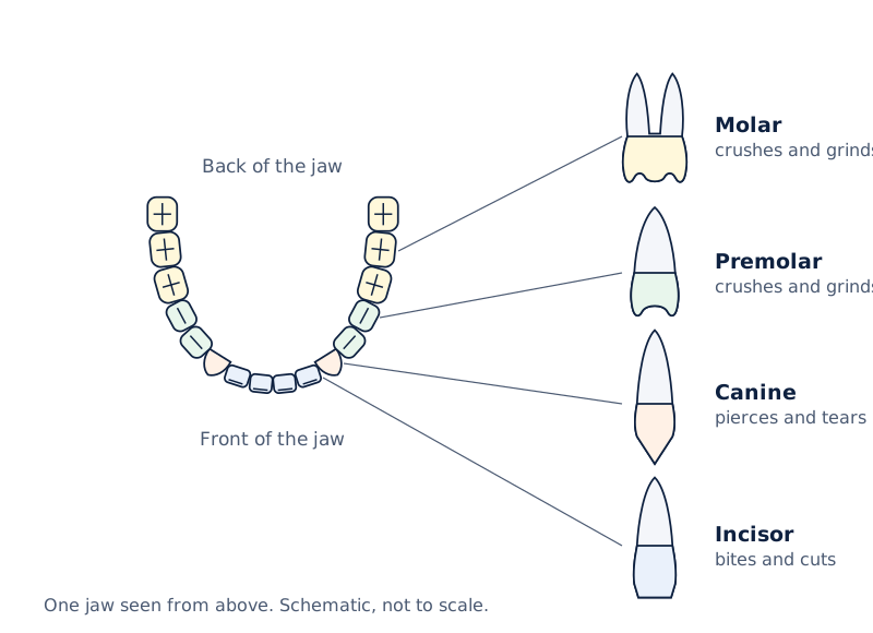
The drawing shows a curved row of teeth, with incisors at the front and broader teeth towards the back. Separate tooth drawings are labelled Incisor, Canine, Premolar and Molar, with lines connecting them to positions in the row.
Drawn by One Young India.
What is the order from the front towards the back?
On either side of a complete permanent jaw, the sequence is incisors, canine, premolars and molars. Starting at the middle of the front of the jaw and moving backwards gives the same order on the left and right sides.
The words cutting, tearing and grinding describe different mechanical actions. They do not name different chemical reactions. Premolars and molars share a grinding role; there is no need to assign chemical digestion to any one tooth type.
When identifying an isolated tooth, connect the visible crown shape with its function. When identifying a tooth in a complete row, use its position as well. A clear explanation combines the name, characteristic shape and main action instead of giving the name alone.
How is the dental formula of an adult written and interpreted?
A dental formula records the numbers of different tooth types on one side of the upper and lower jaws. The standard complete adult human set has two incisors, one canine, two premolars and three molars in each half of each jaw.
In the notation below, I means incisors, C means canines, PM means premolars and M means molars. In each pair separated by a slash, the first number refers to the upper half-jaw and the second to the lower half-jaw on the same side.
Definition: The adult human dental formula is I 2/2, C 1/1, PM 2/2, M 3/3. The slash separates upper and lower counts; it does not ask for division. The order is incisors, canines, premolars, molars.
How does this formula give the complete total?
One half-jaw contains 2 + 1 + 2 + 3 = 8 teeth. The upper jaw has a left half and a right half, so its complete count is 16. The lower jaw also contains 16 in the standard complete set, giving 32 altogether.
| Tooth type | One half-jaw | One whole jaw | Both whole jaws |
|---|---|---|---|
| Incisors | 2 | 4 | 8 |
| Canines | 1 | 2 | 4 |
| Premolars | 2 | 4 | 8 |
| Molars | 3 | 6 | 12 |
| Total | 8 | 16 | 32 |
The table expands the formula by counting both sides and both jaws. For each tooth type, multiply its half-jaw count by four to obtain the full-mouth count. Here the four parts are upper left, upper right, lower left and lower right.
What should be checked before calculating?
First identify whether a stated count describes a half-jaw, one whole jaw or both jaws. Doubling a half-jaw count gives one jaw; doubling that gives both jaws. Confusing these levels changes the total even when the tooth-type counts are correct.
The formula represents a complete permanent dentition. It should not be treated as a promise that every adult has all 32 teeth present. Teeth may be lost due to dental disease, so an observed count can differ from the complete-set count.
Writing only 32 gives the total but loses the distribution among types. Writing the formula preserves that information. A complete interpretation therefore explains what each position means, which parts of the jaws are counted, and how the whole-mouth total follows.
How do milk teeth and permanent teeth differ?
Milk teeth are the first set of teeth, which develops during infancy. Permanent teeth form the later set. The change between these sets is a replacement process, so the teeth present in childhood are not simply a smaller version of the complete adult arrangement.
The complete milk set contains 20 teeth: incisors, canines and molars. It has no premolars. The standard complete permanent set contains 32 teeth and includes premolars as well as incisors, canines and molars. Tooth types and tooth totals must therefore be considered together.
Which comparisons identify the two sets?
| Feature | Milk dentition | Permanent dentition |
|---|---|---|
| Place in the sequence | First set | Later set |
| Complete total | 20 teeth | 32 teeth in the standard complete set |
| Incisors | Present | Present |
| Canines | Present | Present |
| Premolars | Absent | Present |
| Molars | Present | Present |
Diphyodont describes the development of two successive sets of teeth during life. It concerns replacement. Heterodont concerns differences among tooth types within a set. These terms answer different questions and should not be used as substitutes for each other.
Does every permanent tooth replace a milk tooth?
Permanent incisors and canines replace the corresponding milk teeth, and permanent premolars replace milk molars. Permanent molars are added behind the earlier teeth rather than replacing milk teeth. This explains how the later set can contain more teeth than the first set.
Thus, the absence of milk premolars does not mean that the milk set has no grinding teeth. Its molars provide grinding surfaces. Likewise, describing permanent teeth as a replacement set does not mean that each permanent tooth has a milk predecessor of the same name.
Note: Permanent teeth may last throughout life or fall off during old age or due to some dental disease. The word “permanent” names the later set; it does not guarantee that a tooth cannot be lost.
When comparing sets, state the basis of comparison explicitly. Total number, presence of premolars and position in the replacement sequence are separate features. An individual's present count alone is less informative than a description of the complete dentition being discussed.
How should a longitudinal section of a tooth be drawn and read?
A longitudinal section is a view obtained by imagining a cut along the length of a structure. For a tooth, it reveals the relationship between the crown and root, their coverings, and the central pulp space. An outside view cannot show all these internal parts.
Diagram
Longitudinal section of a human tooth
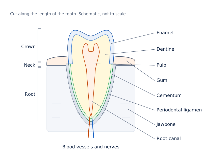
Draw the crown above the gum and the root within a jaw socket. Show enamel over the crown, dentine beneath it, pulp centrally, cementum over the root, and a root canal. Add blood vessels and nerves entering at the root end.
Drawn by One Young India.
Which relationships must the drawing preserve?
- External regions: place crown, neck and root in relation to the gum line, rather than labelling them as layers around the centre.
- Crown covering: show enamel outside the dentine in the exposed crown.
- Root covering: show cementum on the root, with the supporting ligament between it and the jawbone.
- Central structures: show pulp within the cavity and the root canal continuing towards the root end.
A label line for enamel should end on the outer crown layer. A dentine label should end on the hard tissue beneath that layer. A pulp label should identify the central soft tissue, while a pulp-cavity label identifies the space containing it.
How can a diagram support an explanation?
Read from the outside towards the centre when explaining protection and internal structure. Read from the crown towards the root when explaining exposed and embedded regions. These are different directions through the same drawing, and each answers a different structural question.
The blood vessels and nerves belong in the pulp region and its continuation through the root. They should not be drawn as contents of the enamel layer. The root, its cementum covering and the surrounding ligament should also remain distinguishable.
Finish by matching each label to a function: enamel protects the crown, dentine forms the main body, pulp contains living tissue, and the ligament helps anchor the root. This links the drawing with an explanation instead of making it a collection of disconnected names.
How does tooth decay affect the structures of a tooth?
Dental caries, or tooth decay, is the gradual softening and damage of enamel and dentine. Bacteria are microscopic single-celled organisms. Bacteria acting on sugars left in the mouth produce acids, substances that can remove minerals from the hard tooth covering.
Demineralisation means the loss of minerals from a material such as enamel. Dental plaque is the mass of bacterial cells and food particles that sticks to teeth. Plaque covers the surface and prevents saliva from reaching it to neutralise the acid.
Microorganisms are organisms too small to see clearly without magnification; bacteria are one example. Inflammation is a tissue response to injury or infection. Infection occurs when microorganisms invade and multiply in body tissues. Pulp involvement is a possible consequence of untreated decay.
What is the sequence of damage?
- Bacterial cells and food particles accumulate as plaque on the tooth surface.
- Bacteria act on sugars and produce acids; plaque prevents saliva from reaching the covered surface to neutralise them.
- The acids soften or demineralise enamel, beginning damage to the hard outer covering.
- Decay involves the gradual softening of enamel and dentine; if untreated, microorganisms may invade the pulp, causing inflammation and infection.
What the figure shows
Gradual decay of a tooth
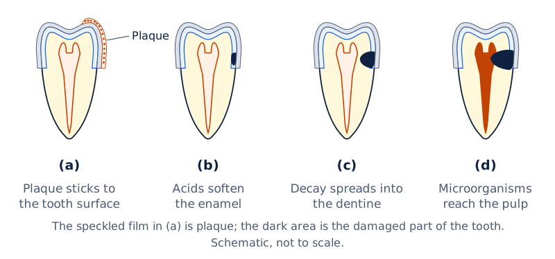
Four small tooth-section drawings are arranged vertically and marked (a), (b), (c) and (d). Across the sequence, the dark damaged region extends progressively further into the tooth. The drawings illustrate progressive damage rather than a labelled map of tooth tissues.
Drawn by One Young India.
How does cleaning interrupt this process?
Brushing removes plaque before bacteria produce acids. Cleaning between teeth removes trapped food particles. Dental floss is a special strong thread moved between teeth for this purpose. Cleaning the teeth and rinsing the mouth after meals help remove material left around them.
Normally bacteria are present in the mouth but are not harmful to us. If teeth and mouth are not cleaned after eating, many harmful bacteria also begin to live and grow there. The presence of bacteria alone is therefore not a complete explanation of decay.
To explain the mechanism, connect sugars, bacteria, acids and tooth damage. Sugar is the material acted on, bacteria produce the acids, and the acids damage the tooth. Preserve the distinction between damage to hard tissues and the possible later invasion of the soft pulp.
Glossary
- Mastication — Chewing food with the teeth to break it mechanically into smaller pieces.
- Crown — The exposed region of a tooth above the gum that acts on food.
- Neck — The region at the gum line between the crown and root of a tooth.
- Root — The embedded region of a tooth accommodated within a socket in the jaw.
- Enamel — The very hard outer covering that protects the crown of a tooth.
- Dentine — Hard tissue beneath the outer coverings that forms most of a tooth.
- Pulp — Soft living tissue within the tooth containing blood vessels and nerves.
- Cementum — The hard covering of the root that provides attachment for supporting fibres.
- Periodontal ligament — Fibrous tissue connecting the tooth root covering to the surrounding jawbone.
- Incisor — A front tooth with a cutting edge used to bite and cut food.
- Canine — A pointed tooth beside the incisors used for piercing and tearing food.
- Premolar — A broad tooth behind the canine that helps crush and grind food.
- Molar — A broad tooth towards the back of the jaw used for crushing and grinding.
- Dental formula — A notation giving tooth-type counts on one side of the upper and lower jaws.
- Dental plaque — A mass of bacterial cells and food particles that adheres to teeth.
Common errors and misconceptions
- Misconception: Crown and enamel mean the same thing. Correct: Crown names the exposed region; enamel names its outer covering. Dentine and the central pulp space lie deeper within the tooth.
- Misconception: Enamel covers the root as well as the crown. Correct: Enamel covers the crown, while cementum covers the root. These coverings occupy different regions.
- Misconception: The pulp cavity is filled with hard dentine. Correct: Dentine surrounds the pulp cavity, which contains soft pulp with blood vessels and nerves.
- Misconception: Canines grind food and molars cut it. Correct: Canines mainly pierce and tear; molars crush and grind. Incisors have cutting edges for biting and cutting.
- Misconception: The numbers 2, 1, 2, 3 describe a whole jaw. Correct: They describe one half-jaw in the complete permanent set. Both halves together contain 16 teeth.
- Misconception: Milk teeth include premolars. Correct: The milk set contains incisors, canines and molars. Permanent premolars replace milk molars; permanent molars are added behind them.
- Misconception: Sugar directly chews away the tooth. Correct: Bacteria act on sugars to produce acids. Those acids soften or demineralise enamel, contributing to decay.
Exam-style questions with model answers
Q1. Distinguish the crown and the root of a human tooth by position. Give one point for each. [2 marks]
- The crown is the exposed region of a tooth situated above the gum line.
- The root is the embedded region of the tooth accommodated in a socket in the jaw.
Q2. State the position and one structural feature of each of these parts of a human tooth: enamel, dentine and pulp. [3 marks]
- Enamel forms the outer covering of the crown, which is the exposed part of the tooth. It is a very hard covering.
- Dentine lies beneath the outer covering and forms most of the tooth. It surrounds the central pulp cavity and extends into the root.
- Pulp occupies the central pulp cavity. It is soft living tissue that contains blood vessels and nerves, rather than a hard outer covering.
Q3. Name the four tooth types in the permanent human dentition. For each type, connect its characteristic crown shape with its main function. [4 marks]
- Incisors have chisel-shaped crowns with cutting edges. At the front of the mouth, they are used for biting and cutting food.
- Canines have pointed crowns. Their pointed form is associated with piercing and tearing food rather than providing a broad grinding surface.
- Premolars have broad crowns with raised points. They are situated behind the canines and help in crushing and grinding food.
- Molars have broad crowns with grinding surfaces. They are situated behind the premolars and are used for crushing and grinding food.
Q4. Each half-jaw of a complete adult human dentition contains two incisors, one canine, two premolars and three molars. There are left and right halves in both upper and lower jaws. Use I for incisors, C for canines, PM for premolars and M for molars. In slash notation, place the upper half-jaw count before the slash and lower count after it. Write the dental formula, then calculate the full-mouth numbers of each of the four types and the total. [6 marks]
- The dental formula is I 2/2, C 1/1, PM 2/2, M 3/3. Each pair gives upper and lower counts on one side.
- There are four half-jaws altogether. Each contains two incisors, so the full-mouth incisor count is 2 × 4 = 8.
- Each of the four half-jaws contains one canine. Therefore, the complete set contains 1 × 4 = 4 canines.
- Each of the four half-jaws contains two premolars. Therefore, the complete set contains 2 × 4 = 8 premolars.
- Each of the four half-jaws contains three molars. Therefore, the complete set contains 3 × 4 = 12 molars.
- Adding the four full-mouth counts gives 8 + 4 + 8 + 12 = 32 teeth in the complete adult dentition described.
Q5. A tooth-section drawing must show enamel, dentine, pulp, cementum and the periodontal ligament. Give one accurate location-and-function statement for each of these five labels. [5 marks]
- Enamel belongs on the outer surface of the crown, above the gum line. This hard covering protects the exposed part of the tooth.
- Dentine lies beneath the outer coverings, surrounding the pulp cavity and extending into the root. It forms the main hard body of the tooth.
- Pulp lies within the central pulp cavity. It contains blood vessels that supply living tissues and nerves involved in sensation within the tooth.
- Cementum covers the root rather than the exposed crown. It provides a surface for the attachment of supporting fibres around the tooth root.
- The periodontal ligament lies between the root covering and the surrounding jawbone. Its fibres help anchor the tooth within its socket in the jaw.
Q6. Give three differences between the complete milk and permanent dentitions in humans, using sequence, total number and the presence of premolars. [3 marks]
- Milk teeth are the first set and develop during infancy. Permanent teeth are the later set that follows the milk dentition during development.
- A complete milk dentition contains 20 teeth. The standard complete permanent dentition contains 32 teeth, so the complete sets differ in total number.
- Milk dentition has no premolars, although it has incisors, canines and molars. Permanent dentition includes premolars as well as those other three types.
Q7. A tooth has plaque containing bacterial cells and food particles, with sugars available. Explain acid production, the effect of plaque on saliva, the damage to enamel, and the possible consequence if untreated decay reaches the pulp. [4 marks]
- Bacteria act on the available sugars and produce acids. This supplies the substances responsible for attacking the tooth's hard covering.
- Plaque covers the tooth surface and prevents saliva from reaching it to neutralise the acid at the covered site.
- The acids soften or demineralise enamel, causing loss of minerals from the protective covering. Tooth decay involves gradual damage to enamel and dentine.
- If decay is untreated, microorganisms may invade the pulp. This can cause inflammation and infection within the tooth's soft living tissue.
Key takeaways
- Teeth break food down mechanically, while salivary amylase changes starch chemically into simpler sugar in the mouth.
- The crown, neck and root are regions of a tooth, identified by their relationship to the gum line.
- Enamel covers the crown, cementum covers the root, and dentine forms most of the tooth beneath these coverings.
- The central pulp contains blood vessels and nerves; supporting fibres around the root help anchor the tooth.
- Incisors cut, canines pierce and tear, and premolars and molars help crush and grind food.
- The adult dental formula counts one side of the upper and lower jaws, giving 32 teeth in the complete set.
- Milk dentition contains no premolars; the later permanent dentition includes premolars as well as incisors, canines and molars.
- Bacteria acting on sugars produce acids that damage enamel; untreated decay may allow microorganisms to invade the pulp.
Test yourself
What does mastication mean, and is it a chemical change?
Mastication means chewing. Teeth mechanically break food into smaller pieces; this action does not itself change the food into a different substance.
Which region of a tooth is at the gum line?
The neck is the region at the gum line, between the exposed crown and the embedded root.
How do the outer coverings of the crown and root differ?
Enamel forms the hard outer covering of the crown, whereas cementum forms the covering of the root.
Why is the pulp different from enamel and dentine?
Pulp is soft living tissue containing blood vessels and nerves. Enamel and dentine are hard parts of the tooth.
What is the tooth-type order from the front towards the back of a permanent half-jaw?
The order is incisors, canine, premolars and molars. The cutting teeth are towards the front and grinding teeth towards the back.
A half-jaw has two incisors, one canine, two premolars and three molars. How many teeth are in both jaws, each with two halves?
One half-jaw contains eight teeth. Both jaws contain four such halves, so the complete total is 8 × 4 = 32 teeth.
Does the absence of premolars mean that milk dentition has no grinding teeth?
No. Milk dentition contains molars, which provide grinding surfaces, even though the set has no premolars.
Which link connects sugar left in the mouth with enamel damage?
Bacteria act on the sugars and produce acids. These acids soften or demineralise enamel and contribute to tooth decay.
Write about it
Turn a question from this chapter into a paper.
The Writing Studio has a structure guide, a rubric and citation tools for 15 formats, from essays to research papers. Drafting is free with an account. Learnacy Essential adds feedback from an AI writing coach and submission to The OYI Review.
Learnacy Essential is ₹1,499 for the year. Publication depends on review and is not guaranteed.