
Cell: The Building Block of Life | CBSE Class 9 Science Notes
On this page

This note covers cells and cell theory, microscopes, the cell membrane, diffusion and osmosis, the cell wall, cell types, cell organelles, mitosis and meiosis, control of cell growth, cell culture and synthetic genetic material.
Why is the cell the building block of life?
A cell is the basic structural and functional unit of life. All living organisms are made of cells. Cells give organisms their structure and carry out the activities that keep them alive, including producing energy, making substances and forming new cells.
Unicellular organisms consist of one cell, as in bacteria and yeast. Multicellular organisms have many cells working together, as in plants, fish, birds and humans. A single-celled organism exchanges materials with its surroundings, while cells in a multicellular organism also interact with neighbouring cells.
A tissue is a group of similar cells performing similar functions. Different tissues form an organ, and several organs work together as an organ system. Even with these levels of organisation, the cell remains the fundamental unit of structure and function.
How did cell theory develop?
In 1665, Robert Hooke examined a thin cork slice with his self-designed microscope. He saw small, box-like compartments and named them cells. His microscope was capable of about 200 to 300 times magnification, meaning enlargement of the object's appearance.
| Scientist | Year | Contribution |
|---|---|---|
| Matthias Schleiden | 1838 | Reported that all plants are made up of cells. |
| Theodor Schwann | 1839 | Found that all animals are made up of cells. |
| Rudolf Virchow | 1855 | Expanded cell theory by stating that new cells form only from pre-existing cells. |
Definition: Classical cell theory has three principles: living organisms consist of one or more cells; cells are the basic units of structure and function; and all cells arise from pre-existing cells.
The theory connects bacteria, plants and animals through their shared cellular organisation. The formation of new cells from existing cells also explains the continuity of life through cell division.
How do microscopes help us study cells?
A cell is usually too small to see with the unaided eye. Resolution is the ability to distinguish two close points as separate. Viewed from about 25 centimetres, written as cm, points about 0.1 millimetre apart can be distinguished by the human eye.
A millimetre, written as mm, is a unit of length. The eye's limit of resolution is 0.1 mm. A microscope makes small structures visible by improving magnification and resolution. Contrast means the difference in brightness between parts of an object.
A light microscope uses visible light and lenses. Its objective lens is near the object, while its eyepiece is the lens through which the image is viewed. Total magnification depends on the magnifying powers of both lenses.
How can the actual size of a cell be estimated?
The field of view is the circular area visible through the eyepiece. A micrometre, written as µm, is one-thousandth of a millimetre: 1 mm = 1000 µm. Measuring this field allows the size of an onion peel cell to be estimated.
- Place a transparent ruler with millimetre markings on the microscope stage, the platform that holds the specimen.
- Focus with the adjustment knob and measure the diameter of the circular field of view in millimetres.
- Convert the diameter to micrometres. A field diameter of 5 mm is 5000 µm.
- Replace the ruler with an onion peel slide and focus on the cells.
- Count the cells along one straight line across the diameter, then divide the field diameter by that number.
The symbol ÷ means division. Estimated cell size = field diameter in micrometres ÷ number of cells along the diameter. For 25 cells across a 5000 µm field, the estimate is 5000 ÷ 25 = 200 µm per cell.
If the eyepiece and objective each magnify 10 times, total magnification is 100 times. The symbol × means multiplication in a calculation and “times” in a magnification such as 10×. The cell's estimated actual size remains 200 µm.
An electron microscope uses a beam of electrons instead of light. It reveals fine cellular details at the nanometre scale. A nanometre, written as nm, is one-billionth of a metre, or 0.000001 mm.
How does the cell membrane control exchange?
The cell membrane, also called the plasma membrane, is a thin boundary surrounding the cell and protecting its contents. It defines the cell as an individual unit. All living cells communicate with their surroundings and neighbouring cells through this membrane.
The membrane is selectively permeable: it allows some substances through while blocking others. This property is central to exchange between a cell and its external environment. The membrane is extremely thin, about 7 to 10 nm thick.
What does the fluid-mosaic model describe?
The fluid-mosaic model describes the membrane's organisation and the movement of its molecules. The membrane contains lipids, meaning fats, and proteins, cellular materials with structural and functional roles. Membrane proteins help substances pass through and act as gatekeepers.
- The lipid bilayer consists of two layers of special fat molecules. Their water-attracting heads face outwards, and their water-repelling tails face inwards.
- Proteins are embedded in the bilayer. Different membrane components together form an arrangement compared with tiles in a mosaic.
- Molecules can move sideways, flip and rotate within the membrane. This movement explains the word “fluid”.
What the figure shows
Cell membrane
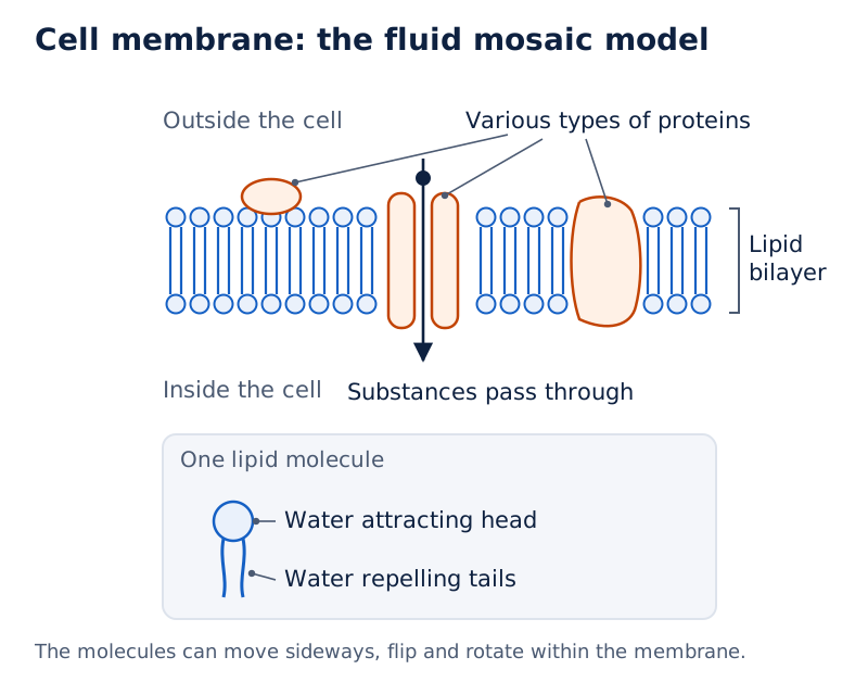
The drawing shows two layers of lipid molecules with their tails directed towards the middle. Larger proteins are embedded among them. The labels identify the lipid bilayer and various types of proteins.
Drawn by One Young India. See Fig. 2.7 in your NCERT textbook
The membrane therefore combines a boundary function with controlled exchange. Its structure helps explain how a cell remains distinct while still interacting with its surroundings. A cell's boundary is an active site of interaction, even in an organism made of a single cell.
How do diffusion and osmosis explain changes in cells?
Concentration describes how much of a substance is present in a given amount of medium. A difference in concentration between two regions is a concentration gradient. Diffusion is the net movement of particles from higher to lower concentration and can occur without a membrane.
A solute is a dissolved substance, such as salt or sugar. A dilute solution contains more water and less solute; a concentrated solution contains less water and more solute. These comparisons help identify the direction of water movement.
Definition: Osmosis is the diffusion of water through a selectively permeable membrane, from a region with more water and less solute towards one with less water and more solute.
Water moves until the concentrations in the two regions become equal. In plants, water enters root cells from the soil by osmosis. The selectively permeable cell membrane is essential to this process.
What does the potato experiment show?
- Carefully cut a potato into two pieces of roughly equal size and record each piece's initial weight using a weighing balance.
- Place one piece in Beaker A containing plain water and the other in Beaker B containing 20 per cent salt or sugar solution.
- Leave the pieces undisturbed for about an hour, or until a visible change in size appears.
- Measure the final weights and compare them with the initial weights to find the change for each piece.
You may observe swelling in Beaker A and shrinking in Beaker B. The potato in plain water gains weight as water enters its cells. The potato in the concentrated solution loses weight as water leaves its cells.
In this experiment, the cell membrane permits water to move in and out but does not allow the salt or sugar molecules through. The observations connect a visible change in the potato with water movement across cellular membranes.
How are surrounding solutions classified?
The extracellular medium is the medium outside a cell; the intracellular medium is inside it. Solution names compare the solute concentration outside the cell with that inside.
| Solution | Outside solute concentration compared with inside | Cell appearance in the comparison |
|---|---|---|
| Isotonic | Equal | Remains similar in size. |
| Hypotonic | Lower | Swells as water enters. |
| Hypertonic | Higher | Shrinks as water leaves. |
Note: Diffusion describes particle movement down a concentration gradient. Osmosis specifically describes water movement across a selectively permeable membrane. A change in cell size in these examples is explained by water movement.
Why do plant cells need a cell wall?
The cell wall is an additional covering outside the cell membrane. It occurs in cells of plants, fungi and bacteria. In general, plants cannot move from place to place, so a rigid covering helps them withstand environmental stresses such as wind and rain.
The wall helps leaves and flowers remain firm, preserves their shapes and supports the plant in an upright position. Although rigid, it is permeable: water and some dissolved minerals can pass through it.
The plant wall is primarily made of cellulose, a carbohydrate, a type of food substance, built from many linked units of the sugar glucose. Cellulose in food acts as roughage, material that helps digestion. The wall's permeability and the membrane's selective permeability together help roots absorb water and nutrients.
How can plant and animal cells be compared under a microscope?
A stain is a colouring material used to make cell structures easier to observe. Safranin and methylene blue are stains used when preparing the plant and cheek cell slides respectively.
- Prepare a temporary slide from a thin onion leaf peel or Rhoeo, also called cradle lily, leaf peel. Mount it with safranin and a coverslip.
- Gently collect cheek cells using a cotton swab or the blunt end of a toothpick, and spread them on a clean slide.
- Add a drop of water and a few drops of methylene blue, then carefully place a coverslip over the cheek cells.
- Observe both slides under a microscope and compare the arrangement of their cells.
Onion or Rhoeo peel cells are box-shaped and regularly arranged. Cheek cells are irregularly arranged. Animal cells such as cheek cells lack a wall, so they can change shape easily. This flexibility supports movement and functioning of animal tissues.
After Rhoeo peel and cheek cells are placed in 20 per cent sugar solution for half an hour, the plant cells retain their outer boundaries. Their contents shrink as the membrane pulls away from the wall. You may observe that the cheek cells have shrunk considerably.
The difference separates two ideas: water leaves both kinds of cell, but the rigid plant wall maintains the outer shape. The cell wall does not prevent the loss of water by osmosis.
How do prokaryotic and eukaryotic cells differ?
Cytoplasm is the semi-fluid, jelly-like material inside a cell. Most cells have a plasma membrane, cytoplasm and a prominent nucleus, the structure containing coded instructions that guide cellular activities. Specialised sub-cellular components are called organelles.
Membrane-bound organelles have their own surrounding membranes. Eukaryotic cells use different organelles to carry out various life processes independently at the same time. Together, these structures build materials, remove wastes and provide energy.
A prokaryotic cell lacks a well-defined nucleus and membrane-bound organelles. A bacterial cell is an example. Most of its cellular activities occur directly in the cytoplasm. Its nucleoid is the region containing genetic material, the information-bearing material involved in inheritance, without a surrounding nuclear membrane.
A eukaryotic cell has a well-defined nucleus and several membrane-bound organelles. Plant and animal cells are examples. The terms refer to the nucleus: “pro” means primitive, “eu” means true and “karyon” means nucleus.
| Characteristics | Prokaryotic cell | Eukaryotic cell |
|---|---|---|
| Primitive nucleus | Present | Absent |
| Diameter of a typical cell | 1 to 10 µm | 10 to 100 µm |
| Number of cells in an organism | Usually unicellular | Can be unicellular or multicellular |
| Membrane-bound organelles | Absent | Present |
| Membrane-bound nucleus | Absent | Present |
What else occurs in the cell interior?
The cytoskeleton is a network of fine fibres in eukaryotic cells. It supports cell structure, maintains shape and enables movement and internal transport. It can be seen as a separate structure only with an electron microscope.
Cell inclusions are stored materials in the cytoplasm. Examples include starch in plant cells and crystals of calcium oxalate or silica in some plant cells. These stored materials are distinct from the specialised structures that perform organelle functions.
What the figure shows
Three kinds of cell
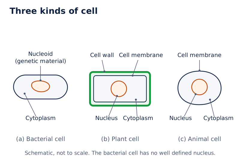
The bacterial drawing labels a nucleoid and cytoplasm. The plant drawing shows a cell wall outside the membrane. The plant and animal drawings both label a nucleus and cytoplasm, showing shared internal features despite their different outer shapes.
Drawn by One Young India. See Fig. 2.10 in your NCERT textbook
How does the nucleus hold genetic instructions?
The nucleus is enclosed by a double-layered nuclear membrane. Its nuclear pores are openings that allow material to move between the nucleus and cytoplasm. This enclosure gives eukaryotic cells their well-defined nucleus.
The nucleolus is the dense, rounded body inside the nucleus. It makes ribosomal subunits, the parts that form ribosomes, which make proteins. One large and one small subunit leave the nucleus and assemble in the cytoplasm.
DNA stands for deoxyribonucleic acid, the molecule carrying genetic information. Genes are functional segments of DNA. Chromosomes consist of DNA and specific proteins and carry information involved in the inheritance of characters from parents to the next generation.
How are chromatin and chromosomes related?
In a non-dividing cell, DNA is part of chromatin, visible as a tangled mass of thread-like material. When a cell is about to divide, this material becomes organised into chromosomes. The chromosomes are visible as rod-shaped structures only when the cell is about to divide.
What the figure shows
Nucleus and genetic material
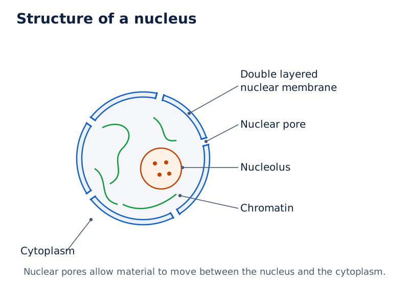
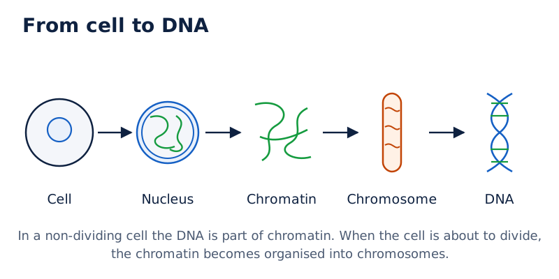
The nucleus drawing labels its double-layered membrane, nuclear pore, nucleolus and chromatin. The accompanying sequence connects a cell and nucleus with chromatin, a chromosome and DNA.
Drawn by One Young India. See Figs. 2.11 and 2.12 in your NCERT textbook
In prokaryotic cells, DNA occurs as a single circular molecule associated with specific proteins in the nucleoid. Absence of a well-defined nucleus therefore does not mean absence of genetic material.
Mature human red blood cells, abbreviated as RBCs, are enucleate, meaning without a nucleus. This leaves more space for haemoglobin, which transports oxygen. They cannot repair or divide themselves and survive for approximately 120 days.
How do organelles make, package and recycle materials?
Protein synthesis means making proteins. Ribosomes may occur freely in the cytoplasm or attached to the endoplasmic reticulum, abbreviated as ER. The ER is a large network within the cytoplasm, continuous with the outer membrane of the nuclear envelope.
The ER helps synthesise and transport proteins, fats and some hormones in some specialised cells. Hormones are substances involved in communication and control in the body. The ER's structure varies with its function.
How do rough and smooth ER compare?
| Feature | Rough endoplasmic reticulum (RER) | Smooth endoplasmic reticulum (SER) |
|---|---|---|
| Ribosomes | Attached to the surface. | Absent from the surface. |
| Appearance | Looks rough under an electron microscope. | Looks smooth. |
| Functions | Mainly protein synthesis and secretion, for example in pancreatic gland cells. | Synthesis and storage of fats and hormones. |
Secretion is the release of substances made by a cell. The Golgi apparatus is a set of stacks of flattened sacs. It modifies, sorts and packages proteins and/or lipids in vesicles, small sacs used to transport materials.
The Golgi apparatus is functionally linked with the ER, cell membrane and other organelles. Its packaged materials may be used in transport, secretion or the formation of lysosomes. Making a substance and preparing it for delivery therefore involve coordinated activities.
What the figure shows
Protein processing and secretion
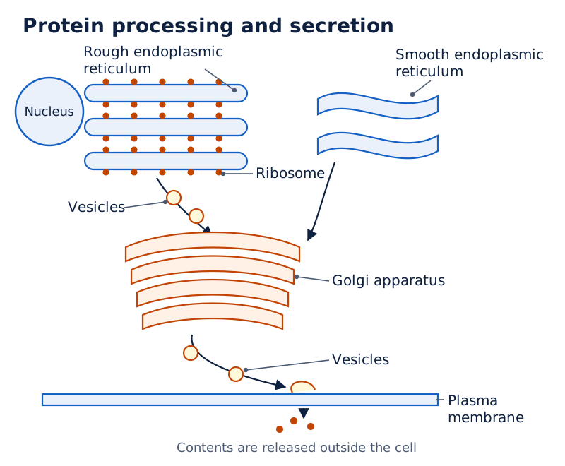
The drawing labels rough ER with ribosomes, smooth ER, the Golgi apparatus, vesicles and plasma membrane. Vesicles are shown between the ER and Golgi stacks and near the membrane where contents are released.
Drawn by One Young India. See Fig. 2.13 in your NCERT textbook
How do lysosomes clean the cell?
Lysosomes are sacs surrounded by a single membrane and filled with enzymes. Enzymes help chemical reactions occur; lysosomal enzymes break down unwanted proteins, carbohydrates, fats and damaged cell parts.
Breakdown products enter the cytoplasm, where they may be reused in other cellular processes. Thus, lysosomes help prevent the accumulation of waste and worn-out organelles while making their breakdown products available to the cell.
Camillo Golgi first observed the Golgi apparatus in 1898 in a barn owl's nerve cells using special staining techniques. Early microscopes could not resolve it clearly, so many doubted its existence. Electron microscope observations confirmed the structure decades later.
How do mitochondria and plastids support cellular activity?
Mitochondria supply energy for most cellular activities and are often called the cell's powerhouses. A single mitochondrion has two membranes. Its outer membrane is smooth and porous, while its inner membrane has finger-like folds called cristae.
Cristae increase the surface area for chemical reactions and facilitate energy production. Cellular respiration is the process in which glucose and other molecules are broken down to release energy. In mitochondria, this energy is stored in adenosine triphosphate, abbreviated as ATP.
ATP acts as the cell's energy currency and is used for most cellular activities. The inner membrane's folds therefore connect the structure of a mitochondrion with its energy-supplying function.
What kinds of plastid occur in plant cells?
Plastids are plant cell organelles involved in food synthesis and storage, with some also providing colour. A pigment is a substance that gives colour. Different plastids contain different pigments or lack them.
| Plastid | Pigment or appearance | Main role |
|---|---|---|
| Chloroplast | Contains green chlorophyll. | Absorbs sunlight for photosynthesis. |
| Chromoplast | Contains pigments other than chlorophyll, which may be yellow, orange or red. | Provides bright colours in flowers and fruits. |
| Leucoplast | Lacks pigment and is colourless. | Stores food such as starch, oils or proteins. |
Photosynthesis is the process by which plants prepare food in sunlight. Chlorophyll, the green pigment in chloroplasts, absorbs sunlight. Each chloroplast has two surrounding membranes and a semi-fluid interior called the stroma.
Disc-shaped membrane structures within the stroma contain chlorophyll. They absorb light energy during photosynthesis. Sugars made in this process are stored in the stroma along with starch granules.
The colours of chromoplasts help attract pollinators, animals involved in the transfer of pollen, and fruit-eating animals that help disperse seeds. Leucoplasts are classified by the food they store. Some in potato and taro, or Colocasia, cells store starch.
What the figure shows
Mitochondrion and chloroplast
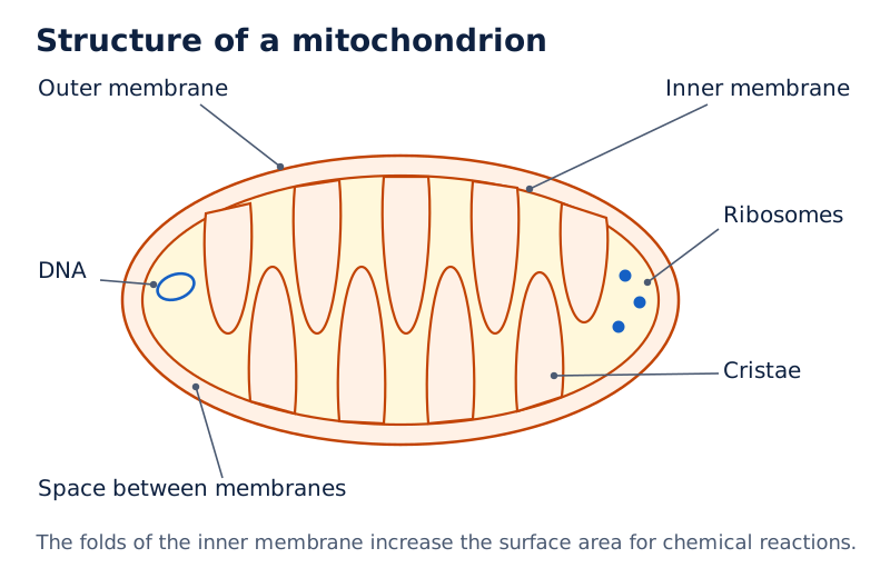
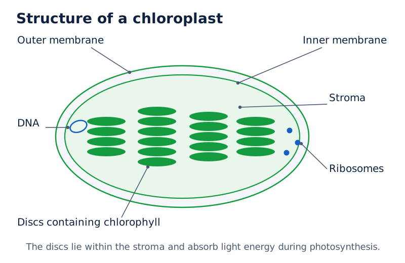
Both drawings label outer and inner membranes, DNA and ribosomes. The mitochondrion shows folded cristae and the space between membranes. The chloroplast drawing labels stroma around internal stacks of discs.
Drawn by One Young India. See Figs. 2.14 and 2.15 in your NCERT textbook
Mitochondria and plastids contain their own DNA and ribosomes, so they can make some of their own proteins. These similarities to certain bacteria suggest a shared evolutionary history with those single-celled organisms.
How do vacuoles support cells, and how do plant and animal cells compare?
A vacuole is an organelle used for storage. In a mature plant cell there is usually one large central vacuole, enclosed by a single selectively permeable membrane. It contains cell sap, a watery fluid holding stored materials.
The vacuole stores water, minerals, sugars and waste material. By holding large amounts of water, it maintains pressure inside the cell and helps keep the plant cell firm. If water is insufficient, the vacuole loses water and the plant wilts as cells become less firm.
Vacuoles are sometimes present in animal cells. They are smaller than plant vacuoles and help temporarily store materials. Plant and animal cells share many organelles, but their coverings, plastids and vacuoles differ.
| Feature | Typical plant cell | Typical animal cell |
|---|---|---|
| Cell membrane | Present. | Present. |
| Cell wall | Present outside the cell membrane. | Absent. |
| Well-defined nucleus | Present. | Present. |
| Mitochondria | Present. | Present. |
| Plastids | Present as special plant organelles. | Absent. |
| Vacuoles | Usually one large central vacuole in a mature cell. | Sometimes present; smaller and used for temporary storage. |
The comparison concerns typical cells. Specialised cells can differ, as mature human RBCs demonstrate. Both plant and animal cells are eukaryotic; possession of a cell wall or plastids does not define whether a cell is eukaryotic.
How do mitosis and meiosis produce new cells?
Cell division forms new cells from pre-existing cells. It supports growth, tissue repair and reproduction. Body growth involves the production of new cells, because individual cells can grow only up to a certain size.
Onion root tips contain continuously dividing cells. Under a microscope, they show different structures corresponding to different stages of division. Prokaryotic and eukaryotic cells both divide, but eukaryotic division follows a more controlled and orderly process called the cell cycle.
What happens during mitosis?
Mitosis produces two genetically identical daughter cells, meaning new cells formed from a parent cell. Each receives the same DNA and chromosome number as the parent. This ensures that genetic information is largely maintained across body cells.
Mitosis supports normal growth, repair, maintenance and asexual reproduction, reproduction without the combination of gametes. Skin cells divide continuously to replace regularly lost cells. Repeated division also allows a single fertilised egg to give rise to the body's trillions of cells.
How does meiosis differ?
Gametes are reproductive cells, such as sperm and eggs, which combine during fertilisation. Meiosis involves two successive divisions and produces four daughter cells, each with half the parent's chromosome number. It is important for sexual reproduction and genetic diversity.
- The parent cell undergoes its first division, forming two daughter cells.
- Each daughter cell has half the chromosome number of the parent cell after this first division.
- A second division, similar to mitosis, occurs in each daughter cell, producing four cells with half the original chromosome number.
- When gametes from two individuals combine during fertilisation, the original chromosome number is restored.
In animals including humans, meiosis occurs only in cells of the testes, the male reproductive organs producing sperm, and ovaries, the female reproductive organs producing eggs. The gametes produced for sexual reproduction create variation, so children resemble their parents without being exactly the same.
| Feature | Mitosis | Meiosis |
|---|---|---|
| Number of daughter cells | Two. | Four. |
| Chromosome number in daughter cells | Same as the parent cell. | Half the parent cell's number. |
| Genetic result | Genetically identical daughter cells. | Variation and diversity associated with sexual reproduction. |
| Importance | Growth, repair, maintenance and asexual reproduction. | Sexual reproduction and creation of genetic diversity. |
Arun Kumar Sharma was an Indian scientist known for chromosome studies and useful laboratory methods for examining plant chromosomes. His work included plant classification, evolution and development.
How are cell growth and cell death controlled?
Normal cells grow and divide in a controlled manner, remain in the appropriate place, perform their functions and eventually die when no longer needed. New cells replace dead cells and perform the same functions. Every cell has a definite lifespan.
In many animal cells, division usually stops when cells contact neighbouring cells. This is contact inhibition. Cancer cells lose this control and continue dividing uncontrollably, which leads to the formation of tumours, masses produced by abnormal cell growth.
Cancerous tumours can invade nearby tissues and spread to other parts of the body, forming new tumours. Plant cells follow a different growth pattern. Because of their rigid walls, they do not show contact inhibition.
Why does controlled cell death matter?
Programmed cell death, abbreviated as PCD, is a genetically regulated, organised process that selectively destroys cells. It is essential for normal development, cellular quality control and immune function, the body's defence against infection.
During development of an embryo, an early stage of a developing organism, PCD removes cells between the digits to help form separate fingers. Without this removal, the hands would be webbed. Cell loss can therefore be part of normal development.
Errors in mitosis lead to uncontrolled division, which can produce tumours and abnormal chromosome numbers in body cells. Errors in meiosis may cause genetic disorders associated with developmental problems or distinctive physical features. Faulty meiosis may also cause early pregnancy loss or reduced fertility.
What do cell culture and synthetic DNA experiments reveal?
Cell culture means growing plant or animal cells outside the body under special conditions. Cells are placed in a nutrient-rich medium that permits growth and multiplication. Suitable temperature, acidic or alkaline conditions, moisture and sterile conditions are maintained.
Sterile conditions prevent contamination by microorganisms. Cell culture helps scientists study cell activity and produce biochemicals, food, medicines and vaccines. The technique depends on providing conditions in which the isolated cells can continue functioning.
What is totipotency?
In 1902, Gottlieb Haberlandt proposed that a living plant cell, even a fully mature one, could develop into a complete plant given suitable nutrients and favourable conditions. Totipotency is this ability to form different kinds of cells and develop into a complete plant.
His proposal laid the foundation for plant tissue culture technology, the field concerned with growing plant cells and tissues under suitable controlled conditions. The nutritional and environmental conditions are essential parts of the idea.
Was a completely new cell made with synthetic DNA?
In 2010, J. Craig Venter and his team studied the complete DNA sequence of the bacterium Mycoplasma mycoides using computer programming. They then chemically synthesised an exact copy of its DNA in the laboratory.
They removed the DNA from a closely related bacterium while retaining the rest of the cell, including its membrane and cytoplasm. After the synthetic DNA was inserted, this cell grew and divided under its instructions, demonstrating DNA's control of cellular structure and activity.
Note: Only the DNA was synthetic in this experiment. The remaining components came from an existing living cell. The scientists did not create a completely new cell from scratch.
Glossary
- Cell — The basic unit that provides living organisms with structure and performs their life functions.
- Resolution — The ability to distinguish two closely positioned points as separate rather than merged.
- Selectively permeable — Allowing some substances to pass through a boundary while preventing others from passing.
- Diffusion — Net movement of particles from higher concentration to lower concentration, even without a membrane.
- Osmosis — Diffusion of water across a selectively permeable membrane from a dilute towards a more concentrated solution.
- Nucleoid — The region of a prokaryotic cell containing genetic material without a surrounding nuclear membrane.
- Organelle — A specialised sub-cellular component that carries out a particular function within the cell.
- Chromatin — Thread-like material containing DNA in a non-dividing cell, which becomes organised into chromosomes before division.
- Gene — A functional segment of DNA, the molecule carrying genetic information in cells.
- Cristae — Folds of the mitochondrial inner membrane that increase surface area for chemical reactions and facilitate energy production.
- Stroma — The semi-fluid material within a chloroplast, surrounding its chlorophyll-containing disc-shaped membrane structures.
- Mitosis — Cell division producing two genetically identical daughter cells with the same chromosome number as the parent.
- Meiosis — Two successive divisions producing four daughter cells with half the chromosome number of the parent.
- Contact inhibition — The stopping of cell division that usually occurs when many animal cells contact neighbouring cells.
- Totipotency — The ability of a living plant cell to develop into a complete plant under suitable conditions.
Common errors and misconceptions
- Misconception: All cells have a well-defined nucleus. Correct: Prokaryotic cells lack one, and mature human red blood cells are also without a nucleus.
- Misconception: The cell wall and cell membrane have identical permeability. Correct: The wall is permeable to water and some dissolved minerals; the membrane selectively permits substances through.
- Misconception: Osmosis means salt moving into a cell. Correct: Osmosis is water movement across a selectively permeable membrane, directed by the difference between the solutions.
- Misconception: A rigid cell wall prevents water loss. Correct: Plant cell contents can shrink in concentrated solution even while the wall preserves the outer shape.
- Misconception: Every plastid is green and performs photosynthesis. Correct: Chloroplasts contain green chlorophyll, chromoplasts contain other pigments and leucoplasts are colourless storage plastids.
- Misconception: Plant cells do not need mitochondria because they contain plastids. Correct: Plant cells have mitochondria, which supply energy, as well as plastids with food-related functions.
- Misconception: Mitosis halves chromosome number. Correct: Mitosis maintains the parental number; meiosis produces daughter cells with half that number.
- Misconception: Synthetic DNA means a whole cell was made from non-living materials. Correct: Venter's team inserted synthetic DNA into a cell whose other components came from a living bacterium.
Exam-style questions with model answers
Q1. State two differences between diffusion and osmosis, considering the substance that moves and the requirement for a membrane. [2 marks]
- Diffusion concerns the net movement of particles from higher to lower concentration; osmosis specifically concerns the movement of water.
- Diffusion can occur without a membrane, whereas osmosis requires a selectively permeable membrane separating the two regions.
Q2. A microscope's field diameter is 5 mm, with 25 onion peel cells along it. Given 1 mm = 1000 µm, estimate a cell's size. The eyepiece and objective each magnify 10×. Find total magnification too. Here mm means millimetre, µm means micrometre and × means times. [3 marks]
- First convert the measured field diameter into micrometres, so that the estimated cell size can be expressed in this smaller unit: 5 × 1000 = 5000 µm.
- Divide the field diameter by the number of cells along it: estimated size of one onion peel cell = 5000 ÷ 25 = 200 µm.
- Multiply the two lens powers: total magnification = 10 × 10 = 100×. The cell appears 100 times larger than its estimated actual size.
Q3. Two roughly equal potato pieces are weighed. One is placed in plain water and one in 20 per cent salt solution. After about an hour, the first swells and gains weight; the second shrinks and loses weight. Explain the observations, naming the process and the membrane property involved. [4 marks]
- The process is osmosis, the diffusion of water across a selectively permeable membrane between solutions of different concentrations.
- The cell membrane is selectively permeable. In this experiment, water passes through it, while the salt does not.
- In plain water, water enters the potato cells from the more dilute surroundings. This explains swelling and the increase in weight.
- In concentrated salt solution, water moves out of the potato cells towards the solution containing less water and more solute. This explains shrinking and weight loss.
Q4. Compare typical prokaryotic and eukaryotic cells under five headings: primitive nucleus, membrane-bound nucleus, membrane-bound organelles, cell diameter and number of cells in the organism. Use µm for micrometre. [5 marks]
- A primitive nucleus is present in a prokaryotic cell and absent in a eukaryotic cell. The prokaryotic genetic region is called the nucleoid.
- A membrane-bound nucleus is absent in a prokaryotic cell. A eukaryotic cell has a well-defined nucleus with genetic material enclosed by a membrane.
- Prokaryotic cells lack membrane-bound organelles, while eukaryotic cells possess them. These organelles enable different life processes to occur independently at the same time.
- The diameter of a typical prokaryotic cell is 1 to 10 µm. The corresponding range for a typical eukaryotic cell is 10 to 100 µm.
- Prokaryotic organisms are usually unicellular. Eukaryotic organisms can be unicellular or multicellular, so having many cells is not a requirement for being eukaryotic.
Q5. Describe five features of a mitochondrion linking its structure with its energy-supplying function. Include its membranes, folds, cellular respiration and the role of adenosine triphosphate (ATP). [5 marks]
- A mitochondrion is surrounded by two membranes. This double covering is an important structural feature of the organelle supplying energy for most cellular activities.
- The outer membrane is smooth and porous, in contrast to the folded inner membrane found within the same organelle.
- The inner membrane forms finger-like projections called cristae. These folds increase the surface area for chemical reactions and facilitate energy production.
- Glucose and other molecules are broken down in mitochondria during cellular respiration. This process releases energy from those molecules.
- The released energy is stored as ATP. ATP acts as the energy currency of the cell and is used for most cellular activities.
Q6. Compare mitosis and meiosis by daughter-cell number, chromosome number, genetic outcome and biological importance. [4 marks]
- Mitosis produces two daughter cells from a parent cell. Meiosis involves two successive divisions that produce four daughter cells.
- Mitosis maintains the chromosome number of the parent cell. Meiosis gives each daughter cell half the parental chromosome number.
- Mitosis produces genetically identical daughter cells. Meiosis is associated with variation and diversity in sexual reproduction.
- Mitosis supports normal growth, repair, maintenance and asexual reproduction. Meiosis is important for sexual reproduction; fertilisation restores the original chromosome number when gametes combine.
Q7. A group of many normal animal cells usually stops dividing on contact with neighbouring cells, but cancer cells continue dividing. Name and explain this control, describe the consequence of its loss, and explain how plant cells differ. [3 marks]
- The control is contact inhibition. In many animal cells, division usually stops when a cell comes into contact with neighbouring cells.
- Cancer cells lose this control and divide uncontrollably, leading to tumours. Cancerous tumours can invade nearby tissues and spread to form new tumours elsewhere.
- Plant cells do not show contact inhibition because of their rigid cell walls. They follow a different pattern of growth from the animal cells described.
Q8. In an experiment, synthetic bacterial DNA was inserted into a related bacterium after its own DNA was removed. Its membrane and cytoplasm were retained, and it then grew and divided. What did this demonstrate, and why was it not a cell made entirely from scratch? [2 marks]
- Growth and division under the inserted DNA's instructions demonstrated that DNA controls cellular structure and activities.
- Only the DNA was synthetic. The membrane, cytoplasm and other retained components came from an already existing living cell.
Key takeaways
- Cells are the structural and functional units of life, and new cells arise from pre-existing cells.
- Microscopes improve visibility through magnification, resolution and contrast; field diameter and cell count allow an estimate of actual cell size.
- The selectively permeable membrane controls exchange, while osmosis explains water movement between cells and surrounding solutions.
- The rigid plant cell wall preserves the outer shape even when cell contents lose water and shrink.
- Prokaryotic cells lack a membrane-bound nucleus and membrane-bound organelles; eukaryotic cells have these internal structures.
- Organelles work together to synthesise, process, transport and recycle materials, while mitochondria provide energy for most cellular activities.
- Mitosis maintains chromosome number in two daughter cells, while meiosis produces four cells with half the parental chromosome number.
- Controlled growth and cell death maintain normal functioning; loss of contact inhibition allows cancer cells to continue dividing uncontrollably.
Test yourself
What three principles make up classical cell theory?
Living organisms consist of one or more cells; cells are the basic structural and functional units; and all cells arise from pre-existing cells.
Why is the membrane model described as fluid and mosaic?
Its molecules can move sideways, flip and rotate, making it fluid. The arrangement of different molecules resembles the tiles of a mosaic.
What is the difference between the nucleolus and the nucleoid?
The nucleolus is a dense body inside the nucleus where ribosomal subunits are made. The nucleoid is the genetic-material-containing region of a prokaryotic cell without a surrounding nuclear membrane.
What causes rough endoplasmic reticulum to look rough?
Ribosomes attached to its surface make rough endoplasmic reticulum appear rough under an electron microscope.
How do chromoplasts differ from leucoplasts?
Chromoplasts contain pigments other than chlorophyll that give bright colours to flowers and fruits. Leucoplasts lack pigments and store food such as starch, oils or proteins.
Why can insufficient water cause a plant to wilt?
The vacuole loses water, reducing the pressure that helps keep the cell firm. The cells become less firm and the plant wilts.
How is the original chromosome number restored after meiosis?
Gametes contain half the parental chromosome number. During fertilisation, gametes from two individuals combine, restoring the original chromosome number.
How does programmed cell death help fingers develop?
It removes cells between the developing digits, allowing separate fingers to form instead of webbed hands.
Write about it
Turn a question from this chapter into a paper.
The Writing Studio has a structure guide, a rubric and citation tools for 15 formats, from essays to research papers. Drafting is free with an account. Learnacy Essential adds feedback from an AI writing coach and submission to The OYI Review.
Learnacy Essential is ₹1,499 for the year. Publication depends on review and is not guaranteed.