
Tissues: Plant and Animal Tissues | ICSE Class 9 Biology Notes
On this page
Watch & explore
Start with a few high-quality watches, then dive into the notes below.
Try an idea before you read. Investigate how specialized plant tissues provide mechanical flexibility and transport nutrients through a 2-step scenario analysis. Explore →
This note covers tissues and division of labour, meristematic and permanent plant tissues, epidermis, supporting and conducting tissues, epithelial and connective animal tissues, muscular and nervous tissues, and the cooperation of tissues in growth, transport, protection and movement.
What is a tissue, and why do multicellular organisms need tissues?
A tissue is a group of cells working together to carry out a particular function. Cells within a simple tissue are similar in structure. In a complex tissue, different kinds of cells cooperate in a shared function.
In a unicellular organism such as Amoeba, one cell performs the functions needed for life. A multicellular organism contains many cells. Different groups of cells become specialised for different tasks, so each group contributes to the functioning of the whole organism.
How does division of labour help?
Division of labour means that different parts perform different jobs. Muscle tissue produces movement, while nervous tissue carries messages. In plants, xylem transports water and minerals, while phloem transports food. Specialisation increases efficiency and allows complex life processes to occur.
These tissues form part of a hierarchy. Cells form tissues; different tissues combine to form organs; organs work together in organ systems; organ systems make up an organism. A tissue is therefore one level of organisation, rather than another name for an organ.
Why do plant and animal tissues differ?
Most plants remain fixed in one place and need support to stay upright. Their cell walls provide strength and rigidity. Animals generally move from place to place, although some, such as sponges, are immobile. Animal cells lack rigid cell walls and can change shape more easily.
Nutrition also creates different requirements. Plants have tissues involved in photosynthesis, whereas animals have tissues involved in processing food obtained from other sources. Both need transport, protection and coordination, but the structures performing these functions differ.
Definition: Specialisation is the development of structural and functional features that enable cells or tissues to perform particular jobs within an organism.
How do meristematic tissues produce growth in plants?
Meristematic tissue consists of actively dividing cells. Their divisions add new cells to the plant body. Growth can increase the length of roots and shoots, increase stem thickness, or allow regrowth after cutting or grazing.
What are meristematic cells like?
These cells are small, with thin walls, dense cytoplasm and a large, prominent nucleus. They are closely packed, with little or no intercellular space. Vacuoles are generally absent. These features are associated with continuous, rapid cell division.
The location of a meristem helps explain the growth it produces. The apical meristem occurs at root and shoot tips. The lateral meristem produces an increase in girth. The intercalary meristem helps grasses regrow after cutting.
| Meristem | Location | Main contribution |
|---|---|---|
| Apical | Tips of roots and shoots | Increase in length |
| Lateral | Along the circumference of the stem | Increase in diameter or girth |
| Intercalary | Base of an internode or just above a node | Regrowth, including growth of grasses after cutting |
A node is a point on a stem where leaves or branches arise. An internode is the portion between two nodes. These terms help locate intercalary meristems accurately instead of placing every growing region at a root or shoot tip.
In a thickening stem, lateral meristematic cells divide and add new cells towards the inside and outside. This increases the stem's diameter. Length and girth therefore describe different kinds of growth, produced by meristems in different positions.
Note: Meristematic tissue is not restricted to the tips of roots and shoots. Lateral and intercalary meristems also contain actively dividing cells.
How does the onion-root investigation demonstrate apical growth?
The growth of onion roots can be investigated by comparing intact roots with roots whose tips have been removed. The important observation is whether each set continues to increase in length. The comparison connects a visible change with the position of actively dividing tissue.
What procedure is followed?
- Fill two glass jars with water and place an onion bulb in each. Identify them as Jar A and Jar B.
- Allow roots to develop and measure their lengths on days 1, 2 and 3.
- On day 3, remove about 1 cm from the root tips of the bulb in Jar B. Leave the root tips in Jar A intact.
- Observe and measure the roots for four more days, recording the lengths from the base of each bulb.
- Compare the change in length in the two jars, particularly after the tips in Jar B have been removed.
What does the comparison show?
The intact roots in Jar A continue growing in length. Roots in Jar B stop growing after their tips are cut. The result supports the conclusion that the root tip contains the apical meristem, whose cells divide actively.
Removing the tip removes the region responsible for continued root elongation in this investigation. The result does not mean that all plant growth occurs at root tips: shoot tips, lateral meristems and intercalary meristems have their own roles.
The useful comparison is the change over time in each jar, rather than a single measurement of final length. Record the root length in each jar on each day, so that the change over time can be compared.
How do permanent tissues develop from meristematic tissues?
Cell division supplies new cells, but those cells do not all remain actively dividing. Some develop features suited to particular functions. The process by which cells become specialised in structure and function is called differentiation.
What happens during differentiation?
- Meristematic cells divide, increasing the number of cells in the plant body.
- Some of the newly formed cells retain their meristematic character and continue dividing.
- Other newly formed cells lose their ability to divide and undergo changes in structure and function.
- These specialised cells form permanent tissues that perform functions such as storage, support or transport.
Permanent tissues are classified as simple or complex according to their cellular composition. Simple permanent tissue contains one type of cell. Complex permanent tissue contains more than one type of cell working together. Complexity here concerns cell types, rather than tissue size.
How do the two classes compare?
| Basis | Simple permanent tissue | Complex permanent tissue |
|---|---|---|
| Cell types | One type of cell | More than one type of cell |
| Examples | Parenchyma, collenchyma and sclerenchyma | Xylem and phloem |
| Functions represented | Storage, photosynthesis, flexibility and support | Transport of materials, with supporting and other associated functions |
| Living cells | Parenchyma and collenchyma have living cells | Xylem includes living parenchyma; phloem is mostly living |
| Dead cells | Most sclerenchyma cells are dead | Xylem conducting elements and fibres are non-living at maturity |
A permanent tissue should not automatically be described as dead. Living parenchyma stores food and can perform photosynthesis in green parts. Conversely, a cell can contribute mechanical strength after death if its walls remain thick and strong, as in sclerenchyma.
Plant tissues also form larger tissue systems. The dermal system forms the covering, the ground system includes the simple supporting tissues, and the vascular system comprises xylem and phloem.
How does the epidermis protect a plant and assist exchange?
The epidermis forms the outermost layer of the plant body. It is made of closely packed cells, generally forming a single layer. Its position allows it to protect underlying tissues from mechanical injury and harmful organisms.
What does the cuticle do?
A waxy layer of cutin, called the cuticle, covers epidermal cells on exposed plant surfaces. It reduces water loss and provides protection. Plants living in very dry habitats may have a particularly thick cuticle, helping them conserve water.
Protection does not mean that the surface prevents all exchange. Different parts of the epidermis are adapted for different functions. Roots absorb water and minerals, while leaves exchange gases and lose water vapour through specialised openings.
How do root hairs and stomata function?
Root hairs are hair-like extensions of root epidermal cells. They increase the surface area available for absorbing water and minerals from the soil. This links the structure of an individual epidermal cell with the plant's water supply.
Stomata are pores in the leaf epidermis that permit gaseous exchange. Water vapour also escapes through them during transpiration. This water loss helps create a pull that contributes to the movement of water through xylem.
Thus, epidermal tissue contributes to protection, absorption and exchange. Its functions depend on where it occurs and the special features of its cells. The waxy covering and root hairs should not be treated as identical adaptations with identical roles.
What the figure shows
Tissues in a sunflower stem
The transverse section and enlarged sector identify the cuticle, epidermis, epidermal hair, collenchyma, parenchyma, sclerenchyma, phloem, lateral meristem and xylem. The drawing places the protective layers outside the internal tissues.
How do parenchyma, collenchyma and sclerenchyma differ?
The three main simple permanent tissues differ in cell-wall structure, living contents and function. Their differences explain why some plant parts store food, some bend easily and others are hard and strong.
What are the features of each tissue?
Parenchyma consists of living, thin-walled cells with intercellular spaces. It mainly stores food. In green plant parts it also performs photosynthesis. Specialised parenchyma in aquatic plants has air spaces that help those plants float.
Collenchyma has living cells whose walls are unevenly thickened at the corners by pectin deposition. It provides support with flexibility. Stems and tendrils can therefore bend without breaking, rather than remaining completely rigid.
Sclerenchyma has thick walls containing lignin. Most of its cells are dead. The strong walls provide hardness and mechanical support. It occurs in stems, leaf veins and hard coverings of seeds and nuts, including coconut husk and walnut shell.
| Feature | Parenchyma | Collenchyma | Sclerenchyma |
|---|---|---|---|
| Cell condition | Living | Living | Mostly dead |
| Wall structure | Thin walls | Uneven thickening at corners | Thick, lignified walls |
| Structural emphasis | Loosely packed cells with spaces | Pectin-rich corner thickening | Hard, strong walls |
| Main roles | Storage and photosynthesis in green parts | Flexible support | Mechanical strength |
| Useful example | Air-containing tissue in aquatic plants | Flexible parts such as stems and tendrils | Coconut husk and walnut shell |
How does wall structure explain function?
Flexible support and rigid support are different requirements. Collenchyma allows bending while providing strength. Lignified sclerenchyma gives hard structures resistance and firmness. Parenchyma's living cells perform functions that require active cellular contents, including storage and photosynthesis.
What the figure shows
Simple permanent tissues
The three panels compare parenchyma with thin walls, collenchyma with conspicuous thickened walls around the cells, and sclerenchyma with thick lignified walls. Compare the wall thickness around the visible cell spaces.
How do xylem and phloem transport materials in plants?
Xylem and phloem are conducting tissues. They are complex permanent tissues because each contains several cell types with related roles. Together they form the vascular tissue system, connecting different parts of the plant through transport.
What does xylem contain?
Xylem transports water and minerals from roots to other parts of the plant. It also contributes strength. Its components are tracheids, vessels, xylem parenchyma and xylem fibres. Tracheids and vessels have tubular forms and thick walls.
Xylem parenchyma is the living component of xylem. The conducting elements and fibres are primarily sclerenchymatous. It is therefore inaccurate to describe the entire tissue simply as living or entirely dead without recognising its different components.
What does phloem contain?
Phloem transports food from leaves to other parts of the plant. Its components are sieve tubes, companion cells, phloem parenchyma and phloem fibres. Unlike xylem, phloem is mostly made of living cells.
Sieve tubes consist of elongated cells joined end to end through perforated walls. Companion cells regulate sieve-tube functions and help with loading and unloading sugars. Phloem parenchyma stores food materials, while phloem fibres provide strength.
| Feature | Xylem | Phloem |
|---|---|---|
| Main material transported | Water and minerals | Food |
| Direction of transport | From roots to other plant parts | From leaves to other plant parts |
| Conducting structures | Tracheids and vessels | Sieve tubes |
| Parenchyma | Living component of the tissue | Stores food materials |
| Fibres | Contribute strength | Support the sieve tubes and provide strength |
| Companion cells | Absent from xylem | Regulate sieve-tube functions |
What the figure shows
Vascular tissues
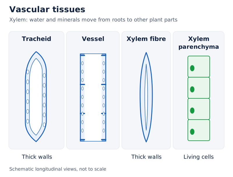
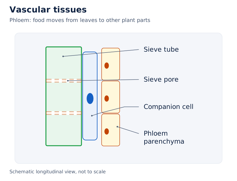
The xylem drawing labels a tracheid, vessel, xylem fibre and xylem parenchyma. The phloem drawing labels a sieve tube, sieve pore, companion cell and phloem parenchyma. The two drawings show different cellular arrangements within conducting tissues.
Drawn by One Young India.
How are epithelial tissues adapted to their functions?
Epithelial tissue covers the body and lines internal structures, including the mouth, lungs, blood vessels and intestine. Its cells fit closely together with very little space between them. This arrangement helps form protective barriers and specialised exchange surfaces.
Why does epithelial structure vary?
A surface involved in rapid exchange has different requirements from one exposed to friction. Thin, flat cells form a short barrier for diffusion. Several layers of closely packed cells provide greater protection to underlying tissues.
Cells involved in secretion produce and release substances. Absorbing cells take up materials from their surroundings. These functions help explain why epithelium includes different cell shapes and arrangements rather than one uniform structure throughout the body.
| Function | Structural feature | Example of location |
|---|---|---|
| Rapid exchange | A single layer of thin, flat cells | Lungs and blood vessels |
| Protection | Several cell layers with tightly packed outer cells | Skin, mouth and oesophagus |
| Secretion | Specialised cells, which may be cuboidal or columnar | Salivary glands, sweat glands and stomach lining |
| Sensory reception | Specialised receptor cells | Nostrils, taste buds and inner ear |
| Absorption | A single layer of tall, pillar-like cells | Small intestine |
How do these tissues contribute to physiological processes?
The thin lining associated with gas exchange in the lungs supports diffusion. The intestinal lining takes up nutrients and water. Glandular epithelial cells release materials such as mucus, enzymes, sweat and saliva. Protective layers reduce injury and entry of microbes.
Identifying an epithelium therefore requires a link between its structure, location and function. Saying that all epithelial tissue is protective misses its roles in absorption, secretion, sensory reception and exchange.
What the figure shows
Epithelial arrangements
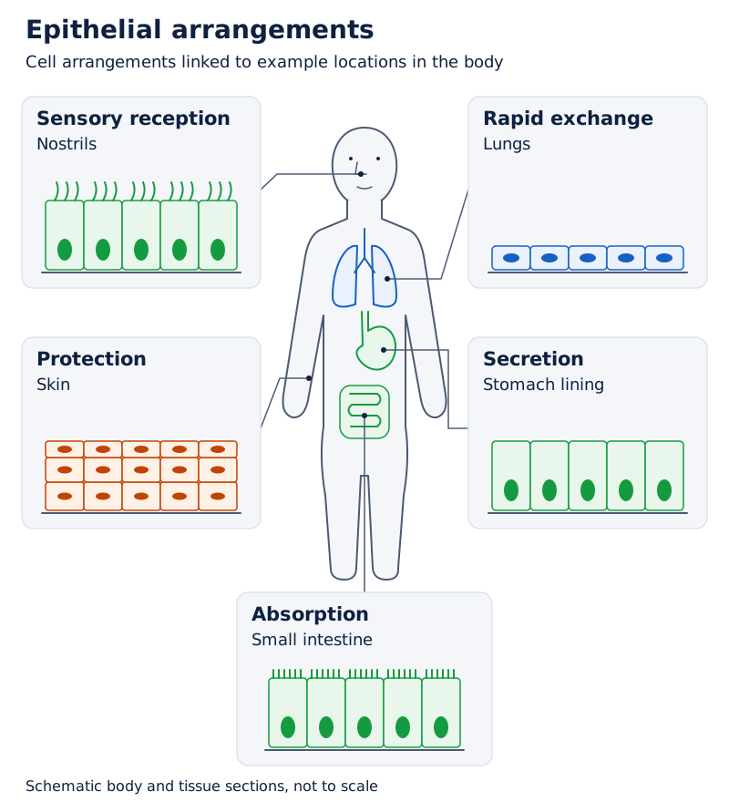
The central body drawing is surrounded by five tissue panels. They show a thin flat layer, a multilayered arrangement, and taller cells with different surface features, illustrating variation among epithelial tissues.
Drawn by One Young India.
Why are blood, bone, cartilage, tendons and ligaments connective tissues?
Connective tissue connects and supports other tissues. Its examples can appear very different: blood is fluid, bone is rigid and cartilage is flexible. These differences reflect the nature of the material surrounding the cells, called the matrix.
How does blood connect body parts?
Blood transports materials between different parts of the body, including nutrients, gases and hormones. Its fluid component is plasma. Red blood cells, white blood cells and platelets are associated with different functions within this transport tissue.
Red blood cells contain haemoglobin, the iron-rich protein responsible for blood's red colour. Platelets help blood clot at an injury. White blood cells collect at infected sites and participate in the body's response to infection.
During exercise, muscles require more oxygen. Breathing becomes faster and blood flow increases. This illustrates cooperation between muscular tissue and the transport function of blood, rather than an isolated action of one tissue.
How do supporting connective tissues compare?
| Tissue | Important feature | Role or connection |
|---|---|---|
| Blood | Fluid matrix | Transport between body parts |
| Bone | Rigid matrix containing calcium and phosphorus compounds | Strength, support and protection |
| Cartilage | Soft, flexible matrix | Cushioning and flexibility, including at bone ends |
| Tendon | Strong band of connective tissue | Connects muscle to bone and transmits pulling force |
| Ligament | Connection between bones | Stabilises joints and limits excessive movement |
Cartilage helps the ear and nose retain a flexible shape and cushions the ends of bones. Bone's mineral-containing matrix provides greater rigidity. Both provide support, but the kind of support differs because their matrices differ.
Tendons and ligaments must be distinguished by what they connect. Tendons transfer a muscle's pull to a bone. Ligaments connect bones to one another, helping maintain joint stability and prevent excessive movement or dislocation.
How do skeletal, smooth and cardiac muscles differ?
Muscular tissue produces movement through contraction. Some movements are under conscious control, such as writing or lifting an object. Others, such as movement of food through the intestine and the heartbeat, occur without conscious control.
Which features identify the three muscle types?
Skeletal muscles are attached to the skeleton and produce voluntary movements. Their fibres are long, cylindrical and unbranched. Each fibre has many nuclei and visible light and dark bands, giving a striated appearance.
Smooth muscles occur in organs such as the stomach and intestine. Their cells are spindle-shaped, have a single nucleus and lack striations. They bring about involuntary movements, including movements associated with digestion.
Cardiac muscle occurs in the heart. Its fibres are cylindrical and branched, with a single nucleus and faint striations. Its rhythmic, involuntary contractions enable the heart to beat throughout life.
| Feature | Skeletal muscle | Smooth muscle | Cardiac muscle |
|---|---|---|---|
| Location | Attached to skeleton | Stomach and intestine | Heart |
| Control | Voluntary | Involuntary | Involuntary |
| Shape | Long, cylindrical, unbranched fibres | Spindle-shaped cells | Cylindrical, branched fibres |
| Nuclei | Many per fibre | One per cell | One per cell |
| Striations | Present | Absent | Present, faint |
| Example of activity | Lifting an object | Movement of food | Heartbeat |
Striations and voluntary control are separate features. Both skeletal and cardiac muscle have striations, but only skeletal muscle is responsible for voluntary movement. The branching of cardiac fibres also distinguishes them from the unbranched fibres of skeletal muscle.
What the figure shows
Three muscle types
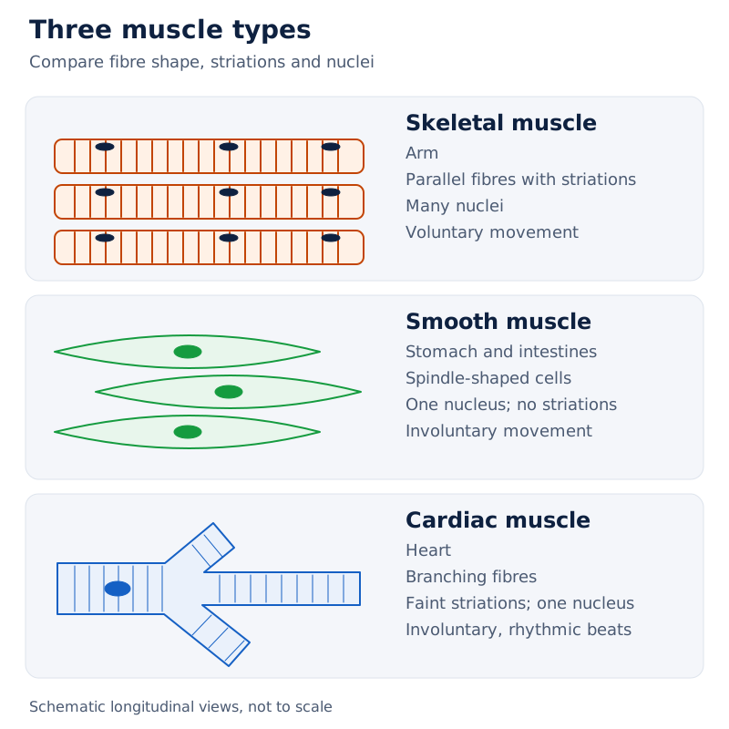
The figure links skeletal muscle to an arm, smooth muscle to digestive organs, and cardiac muscle to the heart. The tissue drawings contrast parallel skeletal fibres, spindle-shaped smooth cells and branching cardiac fibres.
Drawn by One Young India.
How does nervous tissue receive and transmit messages?
Nervous tissue forms the body's communication and coordination network. It helps receive information, process it and bring about responses. Its specialised cells are called neurons, or nerve cells.
What are the main parts of a neuron?
The cell body contains the nucleus and controls the cell's activities. Dendrites receive signals from other neurons. The axon is a long extension that carries messages away from the cell body towards its terminals.
Axon terminals pass messages to other cells. A neuron is therefore structurally suited to communication: it has receiving extensions, a cell body and an elongated conducting part. These parts have different roles within the same cell.
How does nervous tissue cooperate with muscles?
The brain acts as a control centre, coordinating activities and responses across the body. Nervous signals help regulate muscle activity. During exercise, nervous coordination helps the heart beat faster as the body's demand for oxygen rises.
Moving away from something hot is another example of a response involving nervous control. The important tissue distinction is between receiving and transmitting information, performed by nervous tissue, and producing a contraction, performed by muscular tissue.
Communication and movement are therefore related but different functions. A neuron should not be described as a muscle fibre. Nor should the axon be confused with the nucleus: the nucleus lies in the cell body, while the axon extends away from it.
What the figure shows
Structure of a neuron
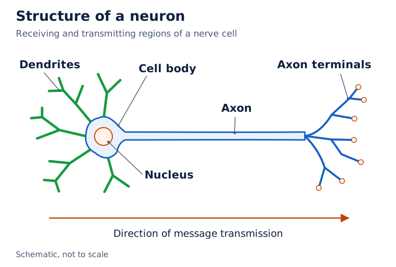
The drawing labels the nucleus within the cell body, the surrounding dendrites, the long axon and the branched axon terminals at its far end. These labels distinguish the receiving and transmitting regions.
Drawn by One Young India.
How do tissues cooperate in plant transport and animal movement?
A physiological process usually involves several tissues working together. The function of one tissue supports the work of another. Water transport in a plant and movement in an animal both demonstrate this cooperation.
How are plant tissues linked in water transport?
Root hairs increase the surface available for absorption of water and minerals. Xylem carries these materials from roots to other parts of the plant. Loss of water vapour through leaf stomata contributes to the pull that helps move water through xylem.
Protective and supporting tissues remain important during this process. The epidermis forms the outer covering, while supporting tissues maintain the plant's form. Phloem supplies a different transport service by distributing food from leaves to other parts.
How does a muscle contraction produce movement?
- Nervous coordination controls activity within the musculoskeletal system.
- A skeletal muscle contracts and exerts a pulling force.
- The tendon joining that muscle to a bone transmits the force to the bone.
- The bone moves at a joint, producing movement of the body part.
- Ligaments help stabilise the joint, while cartilage provides cushioning at the ends of bones.
The musculoskeletal system includes bones, muscles, joints, cartilage, tendons and ligaments. It provides support, helps maintain posture and enables movement. These contributions rely on different tissue properties, including contraction, rigidity, flexibility and resistance to pulling.
A joint is a junction between bones, but the joint alone does not pull the bones into motion. Muscles provide the pulling force. Tendons transmit it, while ligaments and cartilage contribute stability and cushioning. This distinction connects each tissue with its particular role.
Glossary
- Tissue — A group of cells working together to perform a particular function within an organism.
- Division of labour — The sharing of different functions among specialised cells, tissues or other parts of an organism.
- Meristem — Plant tissue containing actively dividing cells that add new cells during growth.
- Differentiation — The process through which cells develop specialised structures and functions.
- Permanent tissue — Plant tissue formed when cells become specialised for functions such as storage, support or transport.
- Parenchyma — Living, thin-walled plant tissue involved in storage and, in green parts, photosynthesis.
- Collenchyma — Living plant tissue with unevenly thickened cell corners that provides flexible support.
- Sclerenchyma — Plant tissue with thick, lignified walls that provides hardness and mechanical strength.
- Xylem — Complex plant tissue that transports water and minerals and also provides strength.
- Phloem — Complex plant tissue that transports food from leaves to other plant parts.
- Epithelium — Closely packed animal tissue forming coverings, linings and surfaces specialised for several functions.
- Matrix — Material surrounding connective-tissue cells, whose properties contribute to the tissue's function.
- Tendon — Connective tissue joining a muscle to a bone and transmitting the muscle's pull.
- Ligament — Connective tissue joining bones to one another and helping to stabilise a joint.
- Neuron — A specialised nerve cell that receives, processes and transmits messages to other cells.
Common errors and misconceptions
- Misconception: All cells in every tissue are identical. Correct: Simple tissues contain one cell type, but complex tissues contain different cell types working together.
- Misconception: Meristems occur only at root and shoot tips. Correct: Lateral and intercalary meristems occur in other positions and contribute to girth and regrowth.
- Misconception: All permanent plant tissues are dead. Correct: Parenchyma and collenchyma are living, and complex tissues also contain living components.
- Misconception: Xylem carries food and phloem carries water. Correct: Xylem transports water and minerals; phloem transports food.
- Misconception: Blood cannot be a tissue because it is liquid. Correct: Blood is connective tissue with a fluid matrix and a transport function.
- Misconception: Tendons join bones to bones. Correct: Tendons join muscles to bones; ligaments join bones to bones.
- Misconception: Every striated muscle is voluntary. Correct: Cardiac muscle is striated but involuntary, whereas skeletal muscle produces voluntary movements.
- Misconception: Joints move bones by contracting. Correct: Muscles contract, tendons transmit their pull, and bones move at joints.
Exam-style questions with model answers
Q1. Define a tissue and state one advantage of division of labour in a multicellular organism. [2 marks]
- A tissue is a group of cells working together to perform a particular function.
- Division of labour increases efficiency because different groups of cells specialise in different tasks, allowing the organism to carry out complex life processes.
Q2. Two onion bulbs are grown in water. Their root lengths are recorded on days 1, 2 and 3. On day 3, about 1 cm is cut from the root tips of bulb B; bulb A remains intact. Afterwards, A's roots continue growing but B's roots stop growing. Identify the tissue involved, give its location and explain the result. [3 marks]
- The tissue involved is the apical meristem, which contains actively dividing cells.
- It occurs at the tips of roots and shoots, where its activity contributes to an increase in length.
- Bulb A retains its growing root tips. Cutting the tips of bulb B removes the actively dividing region, explaining why continued root growth is observed in A but stops in B.
Q3. Compare parenchyma, collenchyma and sclerenchyma in terms of living or dead cells, wall structure and principal functions. Include one example showing the value of flexible or rigid support. [5 marks]
- Parenchyma contains living cells with thin walls and intercellular spaces. It stores food and performs photosynthesis in green plant parts. Specialised forms with air spaces help aquatic plants float.
- Collenchyma also contains living cells, but the walls are unevenly thickened at their corners by pectin deposition. It provides support while allowing plant parts such as stems and tendrils to bend.
- Most sclerenchyma cells are dead and have thick, lignified walls. This tissue provides hardness and strength, as in coconut husk and walnut shell.
- Flexible collenchyma supports bending parts, whereas lignified sclerenchyma suits hard structures. Their different wall properties account for the difference in support.
Q4. Name all four components of xylem and all four components of phloem. State what each tissue transports and explain the function of companion cells. [4 marks]
- Xylem consists of tracheids, vessels, xylem parenchyma and xylem fibres. It transports water and minerals from roots to other plant parts and also provides strength.
- Phloem consists of sieve tubes, companion cells, phloem parenchyma and phloem fibres. It transports food from leaves to other plant parts.
- Companion cells regulate the cellular functions of sieve tubes and help monitor the loading and unloading of sugars.
Q5. Explain how epithelial structure suits rapid exchange in the lungs, protection in the mouth and absorption in the small intestine. [3 marks]
- Rapid exchange in the lungs is supported by a single layer of thin, flat cells, through which substances can diffuse quickly.
- The mouth has several layers of closely packed epithelial cells that protect underlying tissues against friction and mechanical injury.
- The small intestine has a single layer of tall, pillar-like epithelial cells adapted for efficient absorption of nutrients and water.
Q6. Distinguish a tendon from a ligament by the structures each connects and its role. [2 marks]
- A tendon connects muscle to bone and transmits the muscle's pulling force to produce movement.
- A ligament connects bone to bone, stabilises a joint and helps prevent excessive movement.
Q7. Compare skeletal, smooth and cardiac muscle by location, control, fibre shape, nuclei and striations. [5 marks]
- Skeletal muscle is attached to the skeleton and produces voluntary movements. Its fibres are long, cylindrical and unbranched. Each fibre has many nuclei and visible light and dark striations.
- Smooth muscle occurs in organs such as the stomach and intestine and acts involuntarily. Its cells are spindle-shaped, contain one nucleus and lack striations. It contributes to movements involved in digestion.
- Cardiac muscle occurs in the heart and acts involuntarily. Its fibres are cylindrical and branched, contain a single nucleus and have faint striations. Rhythmic contraction produces the heartbeat.
- Striations alone do not establish voluntary control: skeletal and cardiac muscle are both striated, but their control differs.
Q8. Describe the cell body, dendrites and axon of a neuron, and state what axon terminals do. [4 marks]
- The cell body contains the nucleus and controls the cell's activities.
- Dendrites receive signals from other neurons and form the receiving extensions around the cell body.
- The axon is a long fibre that carries messages away from the cell body towards its far end.
- Axon terminals transmit messages to other cells. Together, these parts enable a neuron to receive and pass on information.
Key takeaways
- Tissues organise cells into specialised groups, allowing division of labour and efficient performance of complex life processes.
- Apical, lateral and intercalary meristems contribute to growth in length, growth in girth and regrowth after cutting.
- Parenchyma supports storage and photosynthesis, collenchyma provides flexible support, and sclerenchyma provides hardness and strength.
- Xylem transports water and minerals, while phloem transports food; both contain several types of cooperating cells.
- Epithelial tissues have structures suited to protection, exchange, absorption, secretion and sensory functions in different locations.
- Blood, bone and cartilage have different matrices, while tendons and ligaments differ in the structures they connect.
- Skeletal muscle is voluntary; smooth and cardiac muscles are involuntary, with distinct cell shapes and striation patterns.
- Nervous tissue coordinates responses, while muscles, tendons, bones, ligaments and cartilage cooperate to produce controlled movement.
Test yourself
Which meristem contributes to an increase in stem girth?
The lateral meristem adds cells around the stem and increases its diameter or girth.
What does differentiation mean in the formation of plant tissues?
Cells undergo changes in structure and function, becoming specialised for particular roles such as storage, support or transport.
Why can root hairs improve water absorption?
They increase the surface area of root epidermal cells available for absorbing water and minerals.
Which tissue gives coconut husk its mechanical strength?
Sclerenchyma provides strength through its thick, lignified cell walls; most of its cells are dead.
Why is phloem called a complex permanent tissue?
It contains several cell types, including sieve tubes, companion cells, parenchyma and fibres, which work together.
What is the difference between a tendon and a ligament?
A tendon connects muscle to bone, whereas a ligament connects bone to bone and helps stabilise a joint.
Which muscle is striated but involuntary?
Cardiac muscle is involuntary and has faint striations; it forms the muscular tissue of the heart.
How do dendrites and the axon differ in their roles?
Dendrites receive signals from other neurons, while the axon carries messages away from the cell body towards its terminals.
Try it
ICSE Class 9 Biology: Plant Tissue Adaptations Scenario
Investigate how specialized plant tissues provide mechanical flexibility and transport nutrients through a 2-step scenario analysis.
1A botanist examines a young, non-woody stem that bends during strong wind gusts without snapping. Microscopic cross-sections reveal living, elongated cells with localized, uneven cell wall thickenings of pectin and cellulose at the corners and no intercellular spaces. Which tissue is providing this support?
Correct. The text states that collenchyma is composed of elongated, living cells with localized, uneven cell wall thickenings of pectin and cellulose at the corners, imparting tensile strength and elasticity that allows non-woody stems to bend without snapping.
Incorrect. Sclerenchyma consists of dead cells with uniformly lignified secondary walls that provide rigid reinforcement rather than living cells with corner thickenings.
Incorrect. Aerenchyma is a variant of parenchyma containing large air cavities designed for buoyancy and gas diffusion in aquatic plants, not for mechanical elasticity.
2Further examination of the vascular transport grid reveals living sieve tube cells that have lost their nuclei and vacuoles to lower resistance to nutrient flow. How do these enucleated cells remain functional?
Correct. According to the text, specialized living companion cells with dense cytoplasm and large nuclei are connected to sieve tubes via plasmodesmata, acting as the metabolic engine and genetic brain for the enucleated sieve tube elements.
Incorrect. Phloem fibres are dead sclerenchymatous components that solely provide mechanical strength, not metabolic support.
Incorrect. Xylem vessels are dead conducting tubes responsible for upward water transport, not metabolic or genetic regulation of phloem sieve tubes.
You have successfully analyzed the specialized structural adaptations of plant mechanical and vascular tissues!